
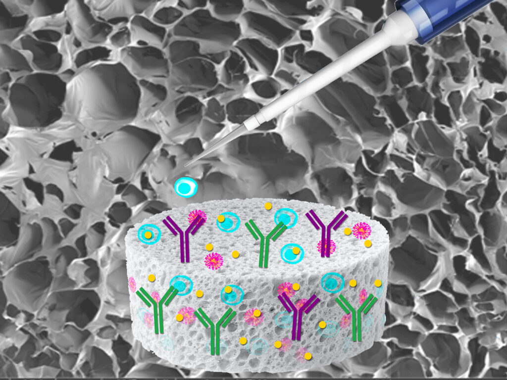
Efficient T-cell engineering is crucial for the success of CAR T-cell therapy research, but it requires multiple labor-intensive steps, including T-cell isolation, activation, and transduction. Traditional activation methods are complex and time-intensive, requiring antibody-coated beads or soluble antibody complexes. Following activation, T cells are then transduced using processes that commonly rely on spinoculation with chemical enhancers (e.g., polybrene), which requires expensive centrifugation equipment, compromises cell viability, and results in varying levels of transduction efficiency.
To overcome the challenges of T-cell engineering using lentiviral transduction, a sponge-like, dissolvable, macroporous alginate—embedded with a blend of activation reagents (rhIL-2, anti-CD3, and anti-CD28 antibodies)—was created and evaluated. The sponge colocalizes the cells and viral particles within the matrix (pore size of 20–300 μm), facilitating efficient transduction and ensuring uniform exposure to activation signals. The cells are then removed from the sponge using a release buffer, ensuring the recovery of transduced cells with minimal handling.
The T-cell sponge was compared against standard T-cell activation methods using two different activation signals and common transduction methods over a series of experiments designed to evaluate activation, transduction efficiency, and cell viability. The sponge was also tested with un-isolated T cells from peripheral blood mononuclear cell (PBMC) samples because PBMCs are increasingly being used for screening purposes in preclinical and clinical research.
To use the T-cell sponge, isolated T cells or PBMCs are combined with the lentivirus, and then directly applied to the T-cell sponge. After 48 hours, the cells are released from the sponge with a buffer for further culture and analysis.
Evaluating T-cell activation
Isolated human primary T cells (4 x 10⁶) were activated using two methods: 1) treatment with a well-known CD3/CD28 T-cell co-activator (conjugate), and 2) using the sponge. The sponge was evaluated at three different multiplicities of infection (MOI 0, 5, 10); the conjugate was evaluated at an MOI of 5. At 48 hours, activation was assessed through CD69 expression using fluorescence-activated cell sorting (FACS). Interferon gamma (IFN-γ) levels were evaluated on days 2 and 4 using a 10-minute lateral flow assay with quantification performed via a smartphone-based system. As a control, T cells that were not exposed to either activation method were also evaluated.
Figure 1 compares the activation signals between the two methods of activation. CD69 expression in T cells activated by the sponge is greater than or similar to those activated by the conjugate. IFN-γ expression was used to further assess T-cell activation, confirming the findings from the CD69 expression data—that the T-cell sponge provides necessary activation signals. Two days after activation, IFN-γ levels were comparable between both activation methods. T cells activated with the sponge have increased IFN-γ expression levels compared to those activated with the conjugate, supporting the use of the sponge as an effective T-cell activation tool. Based on the IFN-γ expression levels, the sponge method promotes greater T-cell activation signals than the conjugate method. Unstimulated control cells show no IFN-γ expression.

Evaluating T-cell transduction
In addition to effective activation, successful preparation of T cells for CAR T-cell therapy research requires efficient transduction and good cell viability following activation.
After evaluation of T-cell activation efficacy with the two methods, transduction efficiency and cell viability were evaluated on days 2, 4, 7, and 9; data for day 11 were also collected for the T-cell sponge method. Transduction efficiency was determined by the percentage of cells that expressed ZsGreen1 after transduction with either spinoculation or the T-cell sponge at an MOI 0 (control), 5, and 10.
To perform the experiment, human primary T cells (4 x 10⁶) were mixed with lentivirus expressing ZsGreen1 at the specified MOIs and exposed to either spinoculation or the T-cell sponge. Spinoculated samples were activated for two days with the conjugate used in the first experiment, exposed to the lentivirus with polybrene, and then centrifuged. The results were analyzed using FACS for ZsGreen1 expression at the specified time points.
For MOIs of 5 and 10 on days 4, 7, and 9, the sponge method performed significantly better than the spinoculation method, even at lower MOIs, as indicated by the higher percentage of T cells expressing ZsGreen1. The cells that were transduced using the sponge method have a peak ZsGreen 1 signal of near 60% on day 7 (Figure 2).

Next, the post-transduction viability for each method was quantified by determining the percentage of cells still living at the specified time points (Day 2, 4, 7, 9, and 11). Cells were evaluated using 7-aminoactinomycin D (7-AAD) staining and FACS analysis. Viability using the sponge method is comparable or better than viability using the spinoculation method, as indicated by the higher percentage of viable cells (Figure 2).
Evaluations from PBMCs
PBMC samples are informative and easily collected from a standard blood draw. After collection, the PBMC layer is separated from other parts of the blood using density gradient centrifugation. To prepare the T cells in this sample for CAR T therapy research, the T cells are typically isolated, activated, and transduced. However, PBMC samples can be applied directly to the T-cell sponge, completely bypassing the isolation step and streamlining activation and transduction into one step for screening applications.
To evaluate the efficacy of the T-cell sponge with PBMC samples, cell subtypes pre- and post-transduction were analyzed (Figure 3). Pre-transduction cell subtypes were analyzed by FACS; post-transduction cell subtypes were analyzed by FACS, and ZsGreen1+ expression was measured. To determine the percentage of cell subtypes post-activation and post-transduction, unstimulated PBMCs (1 x 10⁷) were combined with lentivirus encoding for ZsGreen1 at an MOI of 0 (control), 3, and 12, then directly applied to the T-cell sponge for activation. After 48 hours, the cells were released from the sponge using a release buffer for further culture and analysis.

The data show that the sponge selectively activates and transduces T cells specifically from PBMC samples, potentially saving days in the T-cell engineering and validation workflow.
Conclusions
CAR T-cell therapy research requires engineered T cells. However, traditional methods for producing those cells are laborious, complex, and time-intensive. By using a macroporous alginate-based sponge embedded with rhIL-2, anti-CD3, and anti-CD28 antibodies, researchers can effectively activate and transduce T cells in one step with lower MOIs.
The data indicate that the sponge performs similarly to or better than other methods of activation and transduction by gently fostering colocalization of T cells, activation reagents, and viral particles. This gentle treatment also promotes increased cell viability. Further, by utilizing PBMCs, researchers can bypass a separate T-cell isolation step altogether.
In conclusion, the T-cell sponge is a user-friendly, and effective T-cell activation and transduction tool for T-cell engineering without chemical enhancers or spinoculation from isolated T cells or PBMC samples.
Andrew Farmer, PhD, is the chief scientific officer and head of R&D at Takara Bio USA.