
Sponsored content brought to you by

Biology is complex. To resolve this complexity, researchers need to understand where and what cells are located within tissues, how these cells interact with each other, and how those spatial interactions impact disease progression and patient response. Multiple spatial omic layers are critical to developing this more complete understanding of disease.
The Bruker Spatial Biology portfolio comprises best-in-class solutions for spatial omic layers spanning DNA, RNA, proteins, and post-translational modifications. This breadth and depth reflect a fundamental principle: no single omic layer can fully resolve the biological complexity that drives disease.
High fidelity, high sensitivity, subcellular imaging of the whole transcriptome with proteomics for complete pathway-level spatial insights with the CosMx SMI
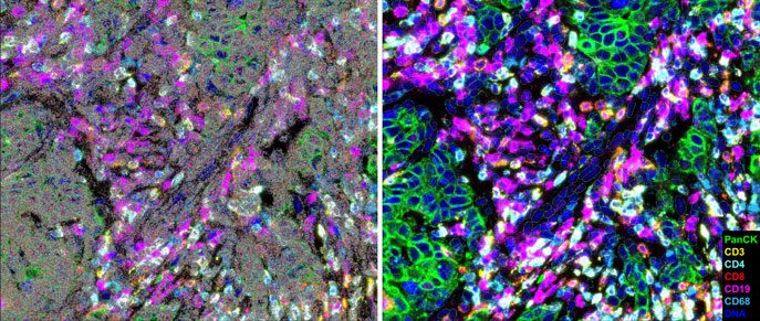
The spatial transcriptomics layer enables high-content discovery, unraveling complex pathways, cell subtypes and states, expression niches, and cell-to-cell interactions. The CosMx® Spatial Molecular Imager (SMI) delivers high sensitivity, subcellular imaging of the whole transcriptome at subcellular resolution, while enabling the simultaneous same-cell multiomic measurement of proteins within the same tissue section. The platform is the first to image the entire protein-coding transcriptome within every single cell at subcellular resolution, enabling researchers to move beyond inferring biology from cell typing alone to directly projecting complete pathway biology in space.
A critical innovation behind the CosMx SMI is its robust direct hybridization chemistry, which does not rely on any enzymatic amplification. This enables whole-transcriptome imaging with superior detection efficiency from FFPE tissues and robust performance across diseases and large studies. In other words, CosMx SMI’s best-in-class sensitivity and specificity results in both the highest transcripts and unique gene counts per cell, which in turn maximizes biological discovery from every single tissue section. Combined with 3D AI-driven cell segmentation, the platform most accurately defines cell boundaries, which is critical for RNA transcript assignment to individual cells and downstream biological analysis.
Moreover, CosMx SMI is the only spatial platform to date to deliver whole transcriptome imaging (>19,000 Human, >21,000 Mouse genes) with high-plex protein imaging (64+ targets) within the same cell. This same-cell multiomic approach enables researchers to reveal pathway–protein spatial relationships, orthogonally validate biomarkers on the same tissue section, and incorporate tissue architecture into their spatial analyses, providing a more complete understanding of tissue biology.
High fidelity, unmatched discovery multiomics to profile from cells to tissue structures for spatial breakthroughs at scale with GeoMx DSP
The GeoMx® Digital Spatial Profiler (DSP) delivers unmatched spatial multiomic discovery at scale through integration of multiple omic layers from pathways to proteins to post-translational modifications. Specifically, the GeoMx DSP combines whole transcriptome regional profiling with >1200 proteins and >130 post-translational modifications from the same tissue section. The result is a mechanistic understanding of spatial biology for the discovery of new biomarkers and signatures at a scale and multiomic breadth not possible with any other spatial approach.
High fidelity, flexible subcellular proteomics for rapid and quantitative spatial phenotyping with the CellScape XR platform
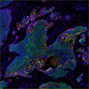
As spatial transcriptomics expands our ability to map gene expression at unprecedented scale, understanding biology ultimately requires connecting transcriptional information to the proteins that execute cellular function and define phenotype. Spatial proteomics therefore provides a key complementary layer of information that can validate and contextualize transcriptomic discoveries while directly measuring the functional state of cells and their microenvironment. Importantly, multiplex immunofluorescence (mIF) also builds naturally on the clinical foundation of immunohistochemistry (IHC), a cornerstone of tissue-based pathology for more than a century, creating a bridge between high-plex discovery and clinically relevant, reproducible tissue assays.
Bruker’s high fidelity CellScape® XR platform overcomes these challenges through integrated advances in instrumentation, assays, and workflow. Leveraging proprietary high dynamic range imaging, the CellScape XR platform enables protein quantification across a continuum of expression. Powered by EpicIF™ technology, the system removes fluorescence signals between staining cycles without harsh antibody stripping, preserving tissue integrity and expanding antibody compatibility for easy assay development and validation.
These advancements directly address the needs of translational and clinical research by increasing throughput, enhancing assay flexibility, and improving signal sensitivity and consistency while maintaining tissue integrity. As spatial biology datasets become increasingly complex, protein expression remains the most informative layer for defining cellular function and phenotype, positioning mIF as a foundational technology rather than simply a validation tool.
As part of Bruker’s broader spatial biology portfolio, the CellScape XR platform provides high-fidelity spatial proteomics that complements transcriptomic and genomic analyses. By delivering rapid, quantitative, and flexible spatial phenotyping at subcellular resolution, the platform helps bridge the gap between biological discovery and clinical impact.
High fidelity performance translates to biological discovery otherwise missed
A recent study1 at Northwestern University provides an example of how these complementary RNA and protein spatial layers can be applied to clinically relevant tissue. Using FFPE samples from patients with ulcerative colitis and Crohn’s disease, the researchers conducted a head-to-head comparison of CosMx SMI and a rolling circle amplification-based platform across several matched tissue blocks, overlapping gene sets, and commercial assays to assess the biological utility and scalability of these platforms for clinical samples.
CosMx SMI delivered superior sensitivity in overlapping genes as measured by transcript counts per cell and unique genes per cell. Importantly, this superiority in sensitivity translated into biological discoveries otherwise missed by the other amplification-based platform. Key immune signals from FOXP3-positive regulatory T cells were confidently detected by CosMx SMI but were not resolved by the other platform under identical conditions, and these findings were subsequently biologically validated.
The study also highlighted the importance of platform robustness when working with real-world FFPE tissue. CosMx SMI yielded superior performance across variable fixation, processing conditions, and multi-site sample handling, supporting its application in large clinical and translational studies where samples are precious, sample quality can vary, and pre-screening workflows are impractical.
Knitting spatial omic layers for capturing disease complexity
The study also demonstrated the value of knitting multiple omic layers together from the same tissue. Specifically, the investigators used an interoperable workflow to generate CellScape and CosMx data from the same tissue section, creating a unified spatial map of proteomic and transcriptomic information. CosMx same-cell multiomics provided an orthogonal validation of the RNA-based findings, demonstrating spatial co-localization of regulatory T cells (Treg) with other immune cells and providing further confidence in RNA-based annotations & biological insights. The CellScape platform revealed a FOXP3 protein expression continuum, resulting in the discovery of Treg subtypes that showed distinct transcriptional profiles, highlighting how quantitative proteomics captures key biological heterogeneity. Together, these results demonstrate the value of integrating quantitative spatial proteomics and high-fidelity spatial transcriptomics to provide a more complete view of disease complexity.
References
1. Li, Y., Wei, C., Yang, W. et al. Spatial transcriptomics atlas of inflammatory bowel disease to guide implementation in research consortiums and clinical trials. Nat Commun 17, 5808 (2026). https://doi.org/10.1038/s41467-026-72482-w

Learn more at brukerspatialbiology.com