
Panelists:
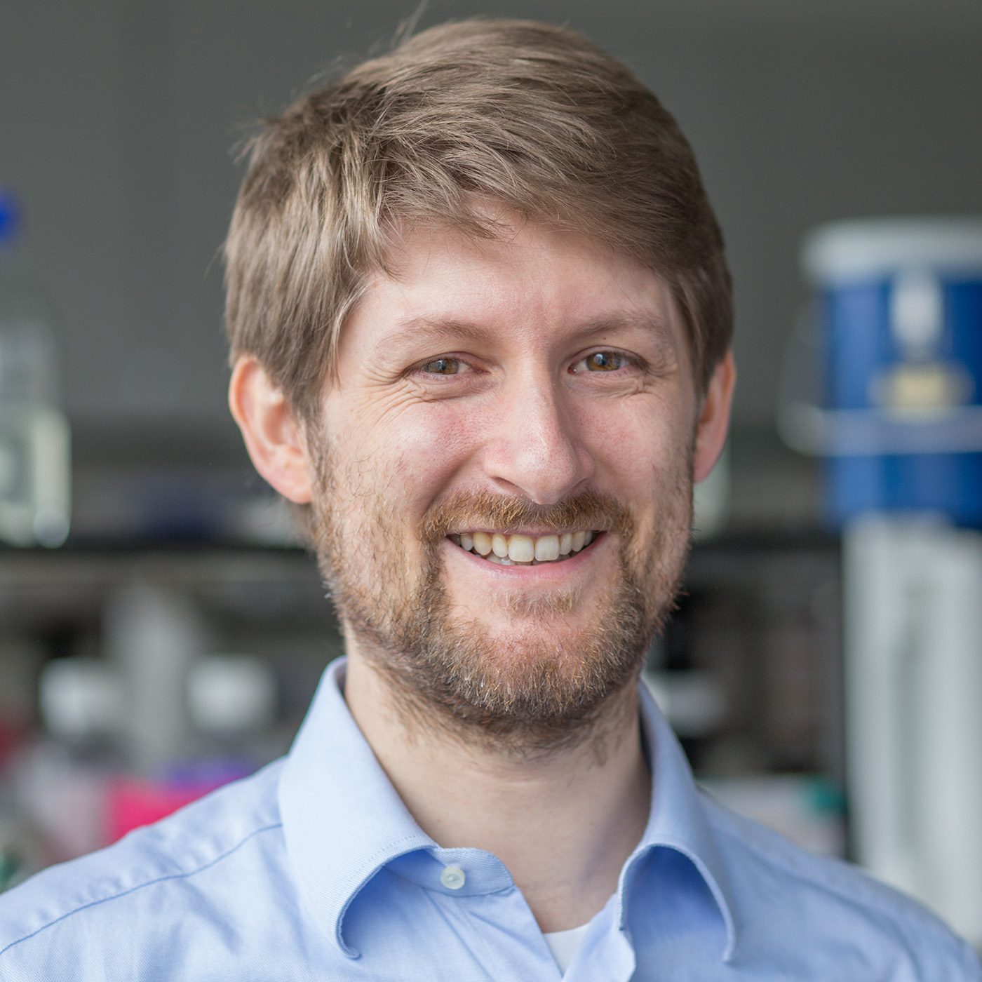
Bernd Bodenmiller, PhD
Associate Professor
Dept. of Quantitative Biomedicine
University of Zürich
ETH Zürich, Switzerland
- Time:
Tumors are complex cellular ecosystems in which cancer cell phenotypes, their spatial organization, and their interactions with immune and stromal compartments determine disease progression and therapy response. To resolve this complexity, the use of highly multiplexed Imaging Mass Cytometry™ (IMC™) together with multiomic profiling and artificial intelligence (AI) can identify clinically relevant features of the tumor microenvironment, providing a rich framework for patient stratification and targeted therapy in precision oncology.
In this GEN webinar, Bernd Bodenmiller, PhD (University of Zürich), illustrates this integrated approach through two recent examples. In a study of 323 epithelial ovarian cancers, tumors with more diverse cell phenotypes and spatial organization recurred faster. His lab also identified a therapy-resistant tumor cell phenotype (TUBB3+/pRB+) associated with recurrence and poor response to standard therapy. Spatial transcriptomics and ex vivo drug screening of 3D sphere models and patient samples showed that these cells resist standard-of-care agents but are sensitive to specific inhibitors. In 498 clear cell renal cell carcinomas, the use of AI revealed three survival ecotypes that predict survival better than basic clinical data and molecular stratification models. Each ecotype has its own genomic and metabolic features and is predictive of treatment outcome. Deep learning predicted the ecotypes directly from routine H&E slides, and the findings were validated across five cohorts comprising more than 2,500 patients.
Webinar attendees will learn how integrating spatial single-cell proteomics by IMC with multiomics and AI identifies therapy-resistant cell populations and prognostic tumor ecosystems. This approach provides a detailed framework for patient stratification and targeted therapy in precision oncology.
A live Q&A session will follow the presentation offering you a chance to pose questions to our expert panelists.
Produced with support from:
