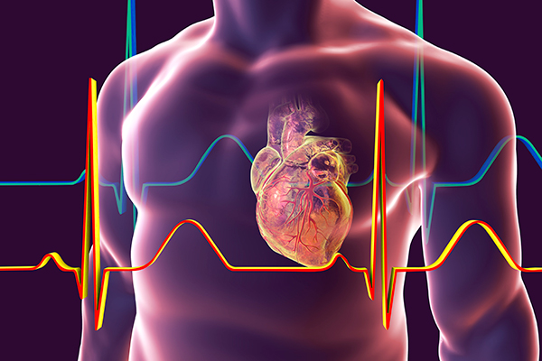
The results of a study demonstrating how circadian rhythms in heart cells are involved in changing heart function over the course of the day could help to explain why shift workers are more vulnerable to heart problems. Led by a team at the MRC Laboratory for Molecular Biology in Cambridge, U.K., in collaboration with AstraZeneca, the findings from rodent studies showed for the first time that heart cells regulate their circadian rhythms through daily changes in the levels of sodium and potassium ions inside the cell.
John O’Neill, from the MRC Laboratory of Molecular Biology, who led the study, said, “Many life-threatening problems with the heart happen at specific times of day, and more often in shift workers. We think that when the circadian clocks in the heart become desynchronized from those in the brain, as during shift work, our cardiovascular system may be less able to deal with the daily stresses of working life. This likely renders the heart more vulnerable to dysfunction.”
O’ Neill and colleagues reported on their in vitro and in vivo research in Nature Communications, in a paper titled “Compensatory ion transport buffers daily protein rhythms to regulate osmotic balance and cellular physiology.”
It is already known there are daily clocks in heart cells, and other tissues are normally synchronized by hormonal signals that align our internal daily rhythms with the day/ night cycle. Daily rhythms of heart function have been recognized for years, and thought to be due to greater stimulation by the nervous system during the day. “Between 6 and 20% of cellular proteins are under circadian control, and the expression of most oscillating proteins peaks during translational “rush hours” that typically coincide with the organism’s habitual active phase,” the authors wrote. Circadian control thus effectively tunes mammalian cell function with daily environmental cycles. The newly reported study has now shown how circadian rhythms within individual heart cells can also affect heart rate.
The different levels of sodium and potassium ions inside and outside heart cells allow the electrical impulse that causes their contraction and drives the heartbeat. It had been thought that cellular ion concentrations were fairly constant, but the new study has demonstrated that heart cells actually alter their internal sodium and potassium levels across the day and night. Such change anticipates the daily demands of our lives, allowing the heart to better accommodate and sustain increased heart rate when we’re active.
The scientists found that these daily rhythms in sodium and potassium occur to allow changes in cellular proteins, with ions literally being pumped out to “make room” for daily increases in protein levels. “We found that dynamic changes in ion abundance drive oscillations in cellular physiology that impart temporal regulation to cardiomyocyte cell function and heart rate,” the authors wrote. “In cultured cells and in tissue we find that compensation involves electroneutral active transport of Na+, K+, and Cl− through differential activity of SLC12A family cotransporters. In cardiomyocytes ex vivo and in vivo, compensatory ion fluxes confer daily variation in electrical activity.”
Study lead author, Alessandra Stangherlin, PhD, was amazed to find that sodium and potassium levels were changing by as much as 30% in isolated cells and heart tissue. This imparts a striking two-fold daily variation to the electrical activity of isolated heart cells. And in mice, this appeared to be just as relevant to understanding daily changes in heart rate as nervous control. “In cardiomyocytes ex vivo and in vivo, compensatory ion fluxes confer daily variation in electrical activity,” the team stated. “Perturbation of soluble protein abundance has commensurate effects on ion composition and cellular function across the circadian cycle … More broadly, our data suggest that the cellular capacity for dynamic ion transport is important for protein homeostasis.”
Understanding how these changes in ion levels alter heart function over the day may help to explain why shift workers might have increased susceptibility to heart problems, because ion rhythms driven by clocks in the heart get “out of sync” with their stimulation from clocks in the brain.
While this study was conducted using cells and mice in the lab, its findings are supported by a recent linked study by collaborators, led by David Bechtold, PhD, at the University of Manchester. Their study demonstrated that circadian rhythms in heart rate and electrical activity are clearly evident in both mice and humans, and that abrupt changes in behavioral routine or sleep patterns can disturb these normal heart rhythms.
Taken together, these studies suggest how lifestyles, such as shift work, which oppose our natural internal clock, may cause internal circadian rhythms within heart cells to uncouple from our behaviors so that heart clocks no longer anticipate the fluctuations in demand that, for most individuals, will be higher in the daytime. This could then feasibly contribute to the increased risk of adverse events, such as arrhythmias and sudden cardiac death, when circadian rhythms are disrupted.
O’Neill said “The ways in which heart function changes around the clock turn out to be more complex than previously thought. The ion gradients that contribute to heart rate vary over the daily cycle. This likely helps the heart cope with increased demands during the day, when changes in activity and cardiac output are much greater than at night, when we normally sleep. It opens up the exciting possibility of more effective treatments for cardiovascular conditions, for example by delivering drugs at the right time of day.”
The findings could have broader implications, beyond cardiovascular function and health, the investigators concluded. “… our data suggest that reciprocal regulation of ion and protein abundance is a ubiquitous cellular mechanism for osmotic homeostasis, which we expect will be of broad relevance to understanding human physiology and disease. For example, since the capacity to buffer cytosolic osmotic potential is reduced when cytosolic ion levels are low, it is tempting to speculate that this may render neural cells more susceptible to protein misfolding and aggregation toward the end of the daily activity cycle, when most mammals normally seek to rest and sleep.”


