
Multiomics analysis combines data from two or more omics layers—such as genomics, transcriptomics, proteomics, and epigenomics. However, published criticisms of multiomics approaches have highlighted concerns about data integration and a tendency toward false-positive findings.1–3
What additional roadblocks might accompany ambitious multiomics projects in 2026? Could any bottlenecks emerge in cost, sample preparation, data integration, or AI implementation? We recently interviewed experts from five companies to gain their perspectives. Here is what they had to say.
Sample preparation
“I sound like a broken record because I can’t stop talking about the importance of sample prep in multiomics work,” says Michael Easterling, PhD, vice president of MALDI/Imaging at Bruker Daltonics.
Easterling stresses challenges such as section-to-section variability, internal standards, FFPE samples, and long sample runs that complicate downstream algorithms. “For multiomics imaging, these issues are not as well defined as some of the bulk methods that have been around much longer,” he notes.
Scott Jelinsky, PhD, senior director of R&D at Thermo Fisher Scientific, agrees. “Different molecular classes have different requirements for sample collection, storage, and preparation,” he says. “Therefore, careful experimental design and standardization are essential.”
Easterling mentions the consequences of inadequate freezing during biopsies, which can disrupt cell structures and lead to ice crystal artifacts. “It is important to really make sure that you have a solid workflow to correctly harvest tissue and provide a good chain of custody from the point that it’s harvested all the way to the analyzer.”
What are some tips for multiomics researchers hoping to prepare their samples in an optimal way? “Community networking is key here,” argues Easterling. “I would say get involved with the overall spatial imaging community and network and find out exactly how to do these things.”
Another practical consideration, according to Jelinsky, is planning the workflow from the beginning so that sample collection, aliquoting, storage, and analytical requirements are compatible across different assays. “Decisions made early in the study can have a significant impact on what can be measured and how confidently the different datasets can be interpreted together,” he notes.
Data integration and analysis
Multiomics technologies generate vast amounts of high-resolution data across modalities that often behave differently from a statistical perspective, notes Najiba Mammadova, PhD, associate director of product management at 10x Genomics. “Multiomics assays have advanced incredibly quickly, and now the analysis and interpretation tools need to keep pace,” she notes.
Mammadova specifically highlights the challenges of computationally aligning multiple omics from different cells or tissues. After all, how can researchers be certain that they are measuring the same cells and biological features?
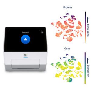
“That’s why I think measuring multiple modalities from the same cell or tissue is so important,” she adds. “It reduces the need to reconstruct the biology after the fact and gives researchers a more direct view of what’s happening in the sample.”
Vanee Pho-Conners, PhD, vice president of global marketing at Mission Bio, agrees that data integration is often a major hurdle with multiomics work. “Each omics layer has its own data structure, distributions, and variabilities, so combining them isn’t plug-and-play,” she explains. Multiomics analysis also often requires cross-disciplinary expertise in biostatistics, machine learning, and biology—capabilities many labs lack in-house.
“It’s important to ask how you can combine these data sets in a clean way where you preserve all the information, reduce the noise, and start to make biological inferences,” adds Easterling. “It’s not something that’s routine today, but the situation is changing rapidly, and new ways to integrate these datasets of varying dimensionality are starting to appear.”
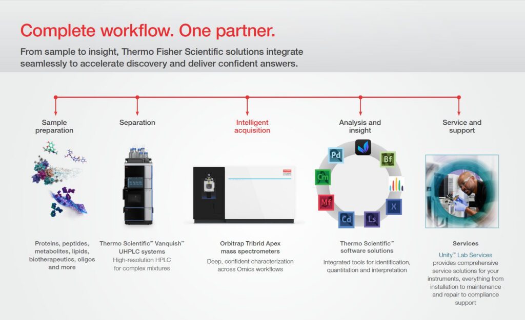
Timing is yet another important consideration, notes Jelinsky. Proteins, RNA, and metabolites may exhibit different kinetics and respond on different timescales, so measurements collected at the same time may not always align. “Understanding those temporal relationships is essential when integrating and interpreting the results,” he says.
AI and multiomics
“I think the rate-limiting multiomics questions are shifting from ‘Can we measure this?’ to ‘Can we interpret it?’ And that’s where AI has enormous potential,” notes Mammadova. “We’re generating increasingly rich, high-dimensional datasets, and AI can help researchers identify patterns, interpret those data more quickly, and ultimately build models that can predict things like disease progression or treatment response.”
However, Easterling warns that even the most advanced AI technologies often cannot overcome challenges associated with inadequate sample preparation. “Unfortunately, if a model’s trained on an inconsistently prepared section, it’ll give you a confident biological story that’s completely wrong. And the reader of this data may have no way to tell.”
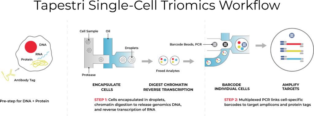
As an example of recent AI advances, Easterling highlights multiomic workflows pioneered at the Medical University of South Carolina for single-cell analysis. The group has developed an AI algorithm that assists lasers in targeting individual cells rather than clusters of cells.4 Pho-Conners also emphasizes how Mission Bio is using machine learning to jointly cluster DNA, RNA, and protein—referred to as triomics—from the same cell. “We are using a combination of triomics and AI-driven integration to establish disease resistance. This really goes beyond correlations using a single omics layer,” she says.
Although Jelinsky is generally optimistic about AI advances in multiomics, he foresees fundamental limitations. “I see AI as a tool that can help researchers navigate complexity and generate better hypotheses rather than as a replacement for scientific judgment. AI does not remove the need for rigorous experimental design or biological validation,” he stresses.
Challenges with extracellular vesicles
Distinct multiomics challenges also exist in analyzing extracellular vesicles (EVs), small membrane spheres released by cells that contain multiple omics (DNA, RNA, protein, metabolites, and lipids).
“EVs are how cells talk to each other, and a living cell sends up to ten thousand a day. This makes them good biomarkers,” says Pierre Arsène, founder and CEO of Mursla Bio.
However, with traditional EV analysis, each potential omics layer is measured from EVs derived from an unknown blend of cells. There is usually no way to know whether a protein signal and an RNA signal came from the same tissue, for instance.
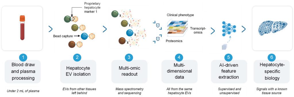
“Tissue-specific extracellular vesicles (TS-EVs) are how we recover that record,” explains Arsène. Mursla’s technology begins with a blood draw. Liver-specific EVs are then captured using antibodies against proprietary surface markers (more specifically, hepatocyte-EV markers). Finally, proteomics and RNA sequencing are performed on two milliliters of plasma for biomarker discovery.
“The great thing about TS-EVs is that they arrive addressed, carrying surface proteins specific to the sending cell and sealed behind a membrane that shields proteins and RNA from the enzymes that would destroy them in blood.”
EvoLiver, Mursla’s blood test for liver cancer surveillance in cirrhotic patients, holds an FDA Breakthrough Device Designation. It analyzes three proteins and five microRNAs derived from hepatocytes. Proteins provide information about tumor surface phenotype and metabolic reprogramming, while microRNAs reveal the regulatory programs driving that phenotype.
As with so many other multiomics platforms, Arsène stresses the importance of TS-EV sample preparation. “Blood handling standards for EVs lag well behind standard liquid biopsy, so our clinical work relied mostly on prospective collection under a common protocol rather than using legacy biobanks.”
The future of multiomics
Mammadova highlights the potential for cost-related challenges in multiomics, particularly as the field moves toward larger studies. “Making these technologies more scalable and cost-effective will be critical to expanding their use across research,” she adds.
“However, I see more overall challenges than roadblocks in multiomics work,” she concludes. “They’re the kind of problems the field works through as technologies mature.”
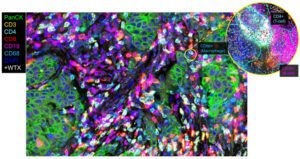
Spatial multiomic layers will also become increasingly critical for decoding the complexity of disease biology, predicts Prajan Divakar, PhD, Bruker Spatial Biology vice president of product management. “Investigators continue to demonstrate the need for more complete spatial datasets, and we are seeing clear applications and momentum for clinical translation,” he says. Divakar highlights Bruker’s CosMx® spatial molecular imager, which measures the whole transcriptome and over 64 proteins.
Finally, Arsène argues that simply adding more layers of analysis without biological resolution is unlikely to advance multiomics research in the future. For instance, in the largest study of a blood-based cancer test ever run (involving around 50,000 participants), incorporating an additional protein layer in advanced adenoma increased the sensitivity only modestly—from about 15% to 16%.5 “More and more layers allow you to gain dimensions, but you end up losing coherence. And no cohort in the world is large enough to train that coherence back computationally.”
Jelinsky agrees that the value of multiomics will lie not in generating more data, but in selecting complementary measurements that address a specific biological question. “I think the future of multiomics will be less about generating the largest possible number of measurements and more about determining which combination of measurements provides the most meaningful biological insight,” he says.
References
- Behrens LMP, Fernandes GDS, Gonçalves GF, et al. Limitations and opportunities in multi-omics integration for neurodevelopmental, neurodegenerative and psychiatric disorders: A systematic review. Neuroscience. 2026;599:76-93. doi: 10.1016/j.neuroscience.2026.01.019.
- Hayes CN, Nakahara H, Ono A, Tsuge M, Oka S. Genes (Basel). From Omics to Multi-Omics: A Review of Advantages and Tradeoffs. 2024;15(12):1551. doi: 10.3390/genes15121551.
- Baião AR, Cai Z, Poulos RC, et al. A technical review of multi-omics data integration methods: from classical statistical to deep generative approaches. Brief Bioinform. 2025;26(4):bbaf355. doi: 10.1093/bib/bbaf355.
- Dressman JW, Bayram MF, Angel PM, Drake RR, Mehta AS. Single-Cell Multiomic MALDI-MSI Analysis of Lipids and N-Glycans through Affinity Array Capture. Anal Chem. 2025;97(24):12493-12502. doi: 10.1021/acs.analchem.4c06233.
- Shaukat A, Burke CA, Chan AT, et al. Clinical Validation of a Circulating Tumor DNA-Based Blood Test to Screen for Colorectal Cancer. JAMA. 2025;334(1):56-63. doi: 10.1001/jama.2025.7515.