
Assessments of DNA misrepair, and the resulting effects on the structural integrity of the genome, are critical for evaluating the safety of cell and gene therapy applications. Most genome editing systems, such as CRISPR-Cas9 systems, are designed to produce precisely targeted double-strand breaks (DSBs), after which the targeted or closely associated sequences are either deactivated or replaced. Unfortunately, no genome editing technology or DNA repair pathway is 100% error-free, and low-frequency but potentially genotoxic structural variants are often observed in parallel with the desired edit(s).
Cell and gene therapy developers need to identify unintended off-target DSBs and consider their consequences, as well as the potential genotoxic consequences of on-target DSBs. Misrepair of DSBs results in the creation of genomic structural variants (SVs), including inversions, deletions, translocations, and complex chromosome aberrations. Drug developers currently use a variety of assay types to detect and identify genomic SVs, including G-band karyotyping, next-generation sequencing, and fluorescence in situ hybridization (FISH).
Directional genomic hybridization (dGH™) is a cytogenomics-based technology uniquely capable of detecting SVs at high resolution on a cell-by-cell basis. The technology, which originated at Colorado State University and came to be developed by KromaTid, is compatible with both targeted and unbiased methods. Besides providing orthogonal confirmation of other testing results, dGH can also identify deleterious gene editing outcomes impossible to assess with conventional detection methods. These include small inversions (as small as 2.0 kb), rearrangements with variable breakpoints and complexity, and the products of genomic instability even when these events are low prevalence, or present in a mosaic or heterogeneous sample.

This tutorial describes two dGH methods for single-cell analysis of batches of edited cells: dGH SCREEN™ for unbiased, whole-genome SV detection, and dGH in-Site™ for targeted detection of on- and off-target SVs and transgene insertions that are either present at a target site or distributed randomly throughout the genome (Table 1). This tutorial maintains that regardless of the degree of heterogeneity in batches of edited cells, pairing dGH SCREEN and dGH in-Site enables measurement of all classes of structural variants, at any genomic location, with industry-leading resolution.
Overview of dGH technology
With dGH, an in situ hybridization technique, unidirectional, single-stranded, fluorescently labeled DNA probes are hybridized to prepared single-stranded metaphase spreads.1,2 The cell preparation technique required to produce metaphase cells with parental-only DNA strands is unique to dGH, but it is straightforward and easily performed in any cell culture–competent laboratory.
The key process steps are as follows:
- Incorporation of a nucleic acid analog (dGH Cell Prep Additive)
throughout a single cycle of DNA replication (S-phase).
2. Mitotic blocking with demecolcine (KaryoMAX® Colcemid™,
Thermo Fisher Scientific) followed by harvest of mitotic cells.
3. Photolytic nicking and removal of the newly synthesized,
singly substituted DNA strands.
The product is a chromosome in which sister chromatids are single-stranded Watson–Crick (5′-to-3′ orientation) complements of each other. As stated previously, dGH probes are single stranded and unidirectional, designed to hybridize to only one parental strand, as seen with standard FISH probes. The combination of fluorescently tagged probes with precisely targeted hybridization enables visualization and quantification of directionally specific genomic rearrangements on individual chromosomes, including inversions (because any change in directionality results in a signal “switch” from one sister chromatid to the other).
dGH in-Site: Analysis of edited cells
In general, dGH in-Site can be used to monitor any specific genomic locus of interest, edited or unedited.3 In the context of gene editing, dGH in-Site is highly valuable for assessing heterogenous global misrepair events in CRISPR-Cas9-edited cell populations. Probes can be designed to target specific gene(s) of interest, including target edit sites and/or inserted transgenes, allowing for the direct assessment of specific loci in a genome.


Figures 1A & 1B show the hybridization results of fluorescently labeled oligonucleotides (green and yellow) designed to bracket a CRISPR-Cas9 edit site on chromosome 19 of normal human CD8 T cells. Targeted dGH can provide information about baseline structural variation of the target site prior to editing for comparison with edited samples to determine the rate of edit-related structural variants and unintended off-site changes.
Figure 1A shows the expected result for a normal cell post-editing, where yellow and green probes are co-localized on each homolog of chromosome 19 (Al, A2). Figure 1B shows that the dGH in-Site assay enables the ready detection of edit-site translocations, resections/deletions, and complex rearrangements (such as chromothripsis). A normal homolog (Bl) with normal signal pattern is evident, as is a reciprocal translocation of the green probe to an off-target chromosome (B2, B3). These likely occur at the cut site on chromosome 19.
dGH SCREEN: Detection of structural variants
The dGH SCREEN assay is composed of individual whole-chromosome paints that collectively target all the human chromosomes. The five-color assay is designed so that identification of all 24 chromosomes is possible through unique combinations of chromosome size, color, and centromere position. An ultra-high-resolution advancement over classic cytogenetic assays such as G-banding and SKY, dGH SCREEN is a next-generation genomics assay for detecting structural variation in metaphase chromosomes. Genomic structural variants can be discovered and tracked on a single-cell basis at unprecedented resolution. The dGH SCREEN assay provides a comprehensive readout of structural variant events by location, type, and incidence in a sample or sample cohort.
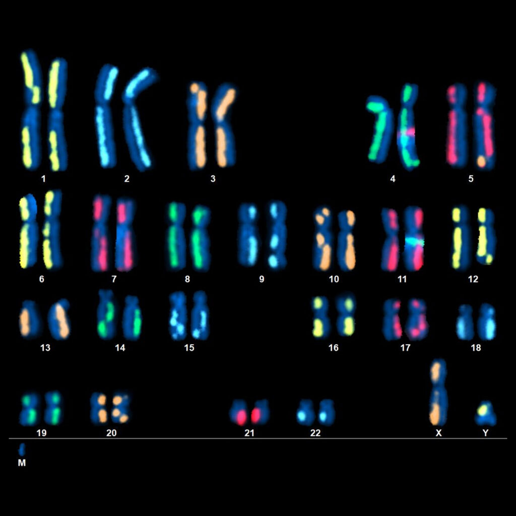
An example of dGH SCREEN is shown in Figure 2. Treated human cells were prepared according to the dGH method,1 then hybridized with the 24 whole-chromosome paints of dGH SCREEN. Among the abnormalities detected were a complex translocation on the q-arm of chromosome 5, a chromatid break on the q-arm of chromosome 9, and an unidentified chromosome fragment (marker). Numerous inversions and sister-chromatid exchanges were also detected, as indicated by the change in position of the paint signal from one chromatid to the other. The assay enables direct detection and identification of all classes of rearrangements, including inversions, at a resolution far superior to that of traditional cytogenetic assays.
Conclusion
Targeted and whole-genome dGH assays provide critical information for cell and gene therapy pipelines. Measuring the formation of potentially deleterious (often cryptic) structural variants is necessary to determine the safest editing approach, to evaluate and release drug product, and to monitor patient health post-treatment. These assays complement each other and can generate a comprehensive dataset around genomic structural changes for process optimization of the nuclease, guide strand, and/or delivery strategies. Importantly, all dGH assays deliver data on a single-cell basis, enabling richly detailed reports on the inherent heterogeneity of structural variation in populations of edited cells.
FDA requirements for testing of genotoxicity and chromosomal aberrations (OECD TG 473) state that such assays must have the ability to detect clastogenicity (for example, deletions, insertions, and translocations due to treatment) and heteroploidy (for example, chromosome loss). Currently available assays that can make low-resolution cell-by-cell measurements include traditional cytogenetic approaches, like G-banding and standard FISH, but in many cases, important edit-associated variants are missed by these methods. The dGH platform can directly detect these variants, including small inversions, along with complex and heterogenous variations often difficult for other assays—even sequencing methods—to identify. dGH assays are ideal for genotoxicity assessment, as well as for informing and optimizing precision medicine strategies based on CRISPR or any other genome editing method.
Erin Cross is vice president, platform, and Christopher Tompkins, PhD, is a member of the scientific advisory board at KromaTiD. Website: www.kromatid.com.
References
1. Robinson E, McKenna MJ, Bedford JS, et al. Directional Genomic Hybridization (dGH) for Detection of Intrachromosomal Rearrangements. Methods Mol. Biol. 2019; 1984: 107–116. DOI: 10.1007/978-1-4939-9432-8_13.
2. McKenna MJ, Robinson E, Taylor L, et al. Chromosome Translocations, Inversions and Telomere Length for Retrospective Biodosimetry on Exposed U.S. Atomic Veterans. Radiat. Res. 2019; 191: 311–322. DOI: 10.1667/RR15240.1.
3. Bailey SM, Cross EM, Kinner-Bibeau L, et al. Monitoring Genomic Structural Rearrangements Resulting from Gene Editing. J. Pers. Med. 2024; 14: 110. DOI: 10.3390/jpm14010110.