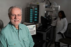
Scientists at Stanford University and the University of California, Irvine, have developed a nanosensor-based test that can diagnose chronic fatigue syndrome (CSF) by measuring how immune system cells in plasma respond to stress. While to date there has been no reliable, standard test for the often-discounted condition—which typically manifests as physical exhaustion and flu-like symptoms, but can be accompanied by a myriad of different symptoms—the newly developed “nanoelectronic assay” correctly identified 20 clinically confirmed cases of CSF, and flagged as negative another 20 healthy controls.
“Too often, this disease is categorized as imaginary,” said Ron Davis, PhD, professor of biochemistry and of genetics at Stanford. “But there is scientific evidence that this disease is not a fabrication of a patient’s mind. We clearly see a difference in the way healthy and chronic fatigue syndrome immune cells process stress.”
Davis, together with Rahim Esfandyarpour, PhD, a former Stanford research associate, and colleagues, reported on the nanoelectronic assay in the Proceedings of the National Academy of Sciences (PNAS) in a paper titled, “A nanoelectronics-blood-based diagnostic biomarker for myalgic encephalomyelitis/chronic fatigue syndrome (ME/CSF).”
CSF, or myalgic encephalomyelitis (ME), is thought to affect more than two million people in the United States, and millions more worldwide. There is no known definitive cause for the condition, but studies have indicated that a combination of factors may be involved, including life stressors, viral or bacterial infections, exposure to toxins, or immune systems disorders. With no single identifiable cause, diagnosing ME/CSF is a matter of ruling out other disorders. “All these different tests would normally guide the doctor toward one illness or another, but for chronic fatigue syndrome patients, the results all come back normal,” Davis commented.
Extensive testing is also a lengthy and costly process, given that candidate CSF/ME patients may report flu-like symptoms, unexplained pain, sensitivity to light, or other stimuli, tingling in different parts of the body, gastrointestinal symptoms or headache, sore throat, intolerance to cold or heat, and other autonomic and endocrine symptoms. “This lag in diagnosis also causes barriers to research, complicating patient recruitment and the handling of heterogeneous samples of patients with only marginally similar conditions,” the authors wrote.
The immune system is a typical focus for ME/CSF research, given that the condition often develops after a viral infection, and patients may have long-term flu-like symptoms. In one recent study patients with ME/CFS showed abnormalities in a third of 63 biochemical pathways measured, “suggesting such metabolic features as potential biomarkers.” Other studies have found that putting stress on peripheral blood mononuclear cells (PBMCs) forces the cells to use up ATP, which is thought to be deficient in ME/CFS patients.

The motivation to investigate PBMCs in plasma was in part driven by the lab’s prior work in severely ill ME/CFS patients, which had shown that patients’ PBMCs behaved very different when in their own plasma, compared with when they were incubated in plasma from healthy individuals.
Esfandyarpour led development of the naonelectronic assay, which effectively measures minute changes in energy as an indicator of immune cell health. “Our assay is designed as an ultrasensitive assay capable of directly measuring biomolecular interactions in real time, at low cost, and in a multiplex format,” the investigators noted.
The device comprises thousands of electrodes that generate an electric current, along with chambers that carry the PBMCs in plasma. In the chambers the immune cells and plasma change the flow of current from one end to another, and this change in electrical activity correlates with sample health.
The test involves adding salt as a stressor to samples taken from either the ME/CSF patients, or healthy control individuals, and comparing how the flow of current is affected. “The assay detects any impedance modulation due to the presence and/or interactions of biomolecules of interest at the active sensing region of the sensors,” the investigators explained.
Large changes in current indicate that the cells are not reacting well to stress. In their tests all of the 20 samples from ME/CSF patients demonstrated clear spikes indicating changes in current, while the 20 control samples generated what would be considered normal results. “We don’t know exactly why the cells and plasma are acting this way, or even what they are doing,” Davis commented. “… we think there might be a correlation between the level of disease severity and signal strength,” the scientists also commented.
The team pointed out that while their initial hypothesis was that the ATP consumption rate would differ in ME/CFS patients’ blood cells compared with healthy control blood cells, the results indicated that salt-stressed samples of PBMC in plasma from the ME/CSF patients, in fact, display “a unique characteristic in their impedance pattern,” which is significantly different to the pattern observed in the control samples. “Our work thus far indicates that the impedance signature itself can potentially represent a distinctive indicator of ME/CFS patients versus controls and can potentially establish a diagnostic metric for the disease,” Davis et al., wrote.
The researchers have in addition applied machine learning tools to their dataset, to develop a classifier for ME/CFS patients, which will be required if the technology is to be used as a robust diagnostic tool. They are now recruiting patients for a larger scale study, to further validate their findings.
“Given the significance of this assay and its reliability, we envision it has the potential to be widely employed in other research laboratories and clinics in the near future as an aid to physicians as well as to our colleagues in the ME/CFS research community,” the authors noted.
The platform is also being adapted to screen for existing FDA-approved drugs that might be repurposed to treat ME/CSF. “Using the nanoelectronics assay, we can add controlled doses of many different potentially therapeutic drugs to patients’ blood samples and run the diagnostic test again,” Esfandyarpour said. The team has already identified one drug candidate that seems to restore immune system cell and plasma function, and which they hope to test further in a clinical trial. “Additionally, we are working on adapting the technology to a platform capable of preclinical testing of drugs and therapies on cells from ME/CFS patients, leading toward development of a portable, handheld, and easy-to-use platform that can be operated by researchers and clinicians at any skill level.”


