Sponsored content brought to you by
![]()
Picture this: you’re a researcher investigating the complex tumor environment of ductal carcinoma in situ (DCIS). Frustratingly, despite decades of technological advancements, why some DCIS cases remain benign while others become invasive carcinoma still isn’t clear.
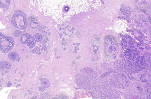
One day, the biopsy pictured here (Figure 1), comes to you along with the opportunity to analyze it with Xenium In Situ, a new subcellular spatial profiling tool from 10x Genomics. This technology provides the unique ability to study tissue biology across molecular, subcellular, cellular, and morphological features to comprehensively characterize cells in their native environment.
You hope that Xenium’s high-resolution, multifaceted view uncovers details about DCIS that other technologies lacking subcellular resolution haven’t been able to pinpoint.
Xenium identifies rare DCIS biology
Hematoxylin and eosin (H&E) assessment—a common technique for clinical
decision making—of this formalin-fixed paraffin-embedded (FFPE-preserved) biopsy determined the sample expresses the human epidermal growth factor receptor 2 (HER2/ERBB2) and estrogen receptor (ER/ESR1) but not the progesterone receptor (PR/PGR).
10x Genomics researchers processed this sample with the 280-gene Xenium Human Breast Gene Panel customized to contain 33 additional genes. The 313 RNA targets were carefully curated from single-cell atlas data for tumorigenic and healthy human breast tissue states (Janesick et al.). Analyte detection and image acquisition were then performed in cycles to generate transcript-specific optical signals and build the subcellular gene expression map.
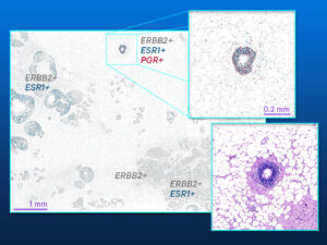
Reviewing the Xenium spatial plot (Figure 2), they discovered that a majority of the analyzed section expresses ERBB2 and ESR1 (HER2/ER double positive) or only ERBB2 (HER2 positive).
Unexpectedly, the Xenium data also identified a small 5.5 mm × 7.5 mm area that expressed ERBB2, ESR1, and PGR (HER2/ER/PR triple positive). Histology missed this unique region, which could impact disease progression or treatment response, underscoring the importance of detecting regions like this, no matter how small.
How was it that Xenium In Situ detected this triple-positive region that histopathology could not?
The Xenium difference: Revealing the unseen and unpredicted
Xenium’s highly sensitive and specific transcript detection, capable of distinguishing between homologous transcripts and detecting lowly expressed transcripts of interest, was particularly important for the identification of the triple-positive region. Because Xenium performed transcript profiling across the entire tissue section, 42 mm2 for this sample, the triple-positive region, measuring a mere 0.1 mm2, was also profiled. Some spatial technologies limit profiling to preselected regions of interest chosen before the experiment. Such a sparse region could have been easily overlooked since its unique biology was not identified via traditional methods.
See your tissues like never before
Xenium In Situ delivers the power of single-molecule RNA detection and spatial mapping for up to thousands of RNA targets at subcellular resolution in fresh-frozen or FFPE tissues. In addition to spatial gene expression, Xenium offers:
- The capability to visualize multiplexed protein detection alongside RNA transcript maps.
- A nondestructive assay that allows seamless incorporation of H&E and immunofluorescence analysis of the same tissue section post-Xenium runs.
- Immediate access to insights with exploration-ready data the moment your run is done.
Whether you are investigating the intricacies of cancer, neurodegenerative disease, immune response, or beyond, high-resolution subcellular profiling with Xenium In Situ can help you uncover new insights.
Reference
Janesick et al. High resolution mapping of the breast cancer tumor microenvironment using integrated single cell, spatial and in situ analysis of FFPE tissue. bioRxiv (2022). doi: 10.1101/2022.10.06.510405.

Discover how Xenium In Situ can transform your research 10xgen.com/xenium-gen

