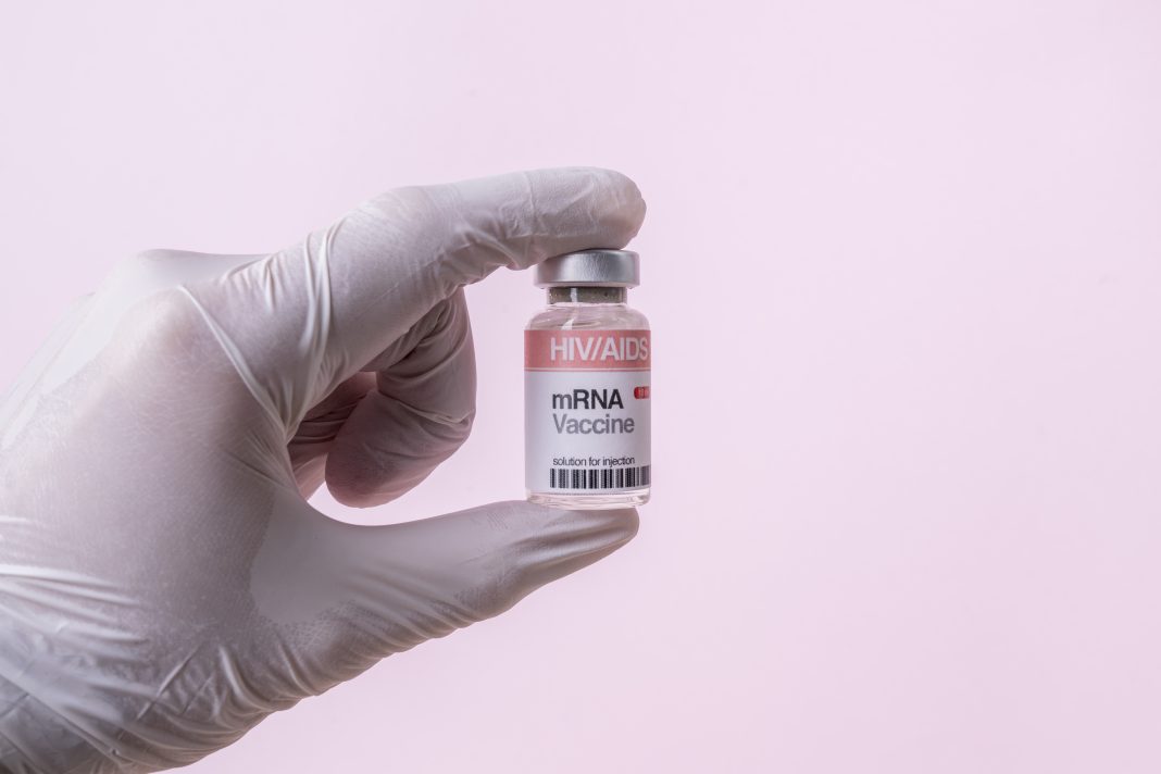
A well-known technique for separating polymers, such as proteins, has been adjusted for analysis of mRNA-lipid nanoparticle (LNP) vaccines. Multidetector Field Flow Fractionation (MD-FFF) has been in use in mRNA research for several years. But it has seen a huge growth of interest for mRNA-LNP manufacturing, according to Jérémie Parot, PhD, a research scientist at SINTEF (Norway), a large European research organization.
According to Parot, whose colleague Sven Even Borgos, PhD, is due to present on MD-FFF for mRNA-LNP at the Bioprocessing Summit in Boston in August, “On the industry side, there’s been a huge boom since COVID started. There are papers from FFF users highlighting its incredible potential and as soon as pharmaceutical companies realized it was super-powerful, they started using it for RNA-LNPs.”
SINTEF is using MD-FFF to analyze samples for pharmaceutical clients, according to Parot, and the systems they’re using for RNA-LNP analysis are different from traditional MD-FFF. “The technique is well-known for nanoparticles, or proteins, but LNPs are different and fairly fragile, so instrument manufacturers needed to develop a new separation device called frit-inlet channel that has less effect on particles,” he explains
MD-FFF is a liquid-based separation technique. Compared to dynamic light scattering techniques, MD-FFF gives a high resolution because it physically separates particles into different sized buckets before they are analyzed.
“You can tell if you’ve got a big population of particles, and a tiny one, or several different-sized populations,” says Parot.
The MD-FFF process involves injecting a stream of liquid into a channel with a membrane at the bottom. The liquid undergoes two separation phases. First, being pushed downwards under pressure towards the membrane, then being allowed to relax and spread out through the channel, which sorts them by “size.”
After the particles have been separated, they can have important features measured using various detectors such as UV/Vis, multi-angle light scattering or by studying their refractive index. Parot says the technique gives a high-resolution analysis while not damaging the fragile LNPs.
“Due to LNPs being a single layer of lipids, we were destroying a significant part of the sample, so a manufacturer came up with a channel that’s gentler,” he continues. “It has a shorter, less strong [first] step, but keeps the diffusion, relaxing step and, from our investigations, that is working really well.”


