
Sponsored content brought to you by

Biological systems are inherently three-dimensional—tissues form intricate layers, networks, and architectures where cells interact in ways that extend far beyond a flat plane. To capture the true complexity of biology, analysis must reflect this dimensionality, providing both breadth and depth across intact tissue structures.
Traditional single-cell and spatial transcriptomics approaches in thin tissue sections have provided powerful insights into gene expression and cellular organization. Yet, by their very nature, these methods capture only a portion of the story. The Pyxa system from Stellaromics expands upon these foundations by enabling spatial transcriptomics in intact thick tissues, opening an entirely new dimension of discovery.
 Your 3D spatial transcriptomics solution
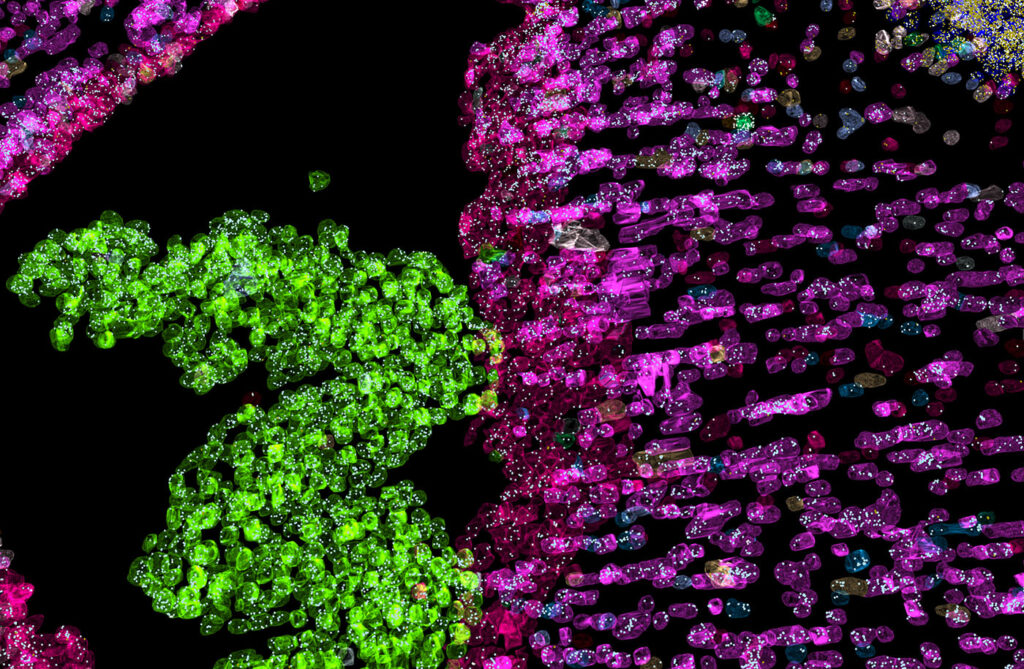
Pyxa is purpose-built for analyzing spatial transcriptomics from intact thick tissue sections in three dimensions. Pyxa leverages the innovative STARmap technology to enable RNA transcript detection with subcellular resolution within issue sections up to 100 microns. The Pyxa system delivers a complete end-to-end process: streamlined reagent kits for simplified sample preparation, automated high-speed imaging for rapid data acquisition, integrated computational analysis, and intuitive visualization tools.
eXtending into the third dimension
Traditional spatial transcriptomic approaches in thin tissue slices cannot capture the full scope of cell-cell interactions and tissue architecture as it exists in situ, which limits our ability to truly understand biological systems. Large cells such as neurons and structures such as blood and lymphatic vessels are not fully captured in thin tissue sections, nor is tissue anatomy such as cortical layers in the brain or the internal structure of the kidney. And reconstructing 2D sections into a 3D approximation is error-prone, introducing misalignments and losing cellular context between serial sections.
With Pyxa, researchers can finally see what was previously invisible.
3D spatial transcriptomics overcomes these limitations by examining intact tissue volumes that preserve their natural architecture. Scientists can now visualize how cells are positioned relative to each other and within the overall architecture of the tissue, revealing anatomical networks like blood vessels or neural circuits. Because Pyxa resolves transcript localization at the subcellular level, this technology can also be used to identify rare cell populations or novel gene expression patterns that define unique cell subsets.
Applications in Disease Research and Drug Development
3D spatial transcriptomics has already been used to make critical discoveries that were previously not possible using existing spatial transcriptomics platforms.
In neuroscience, 3D spatial transcriptomics has been used for atlas creation across entire neural networks. The 3D perspective was particularly critical for mapping neuronal connectomics, where rare yet crucial cell-to-cell interactions often missed in 2D tissue slices could be detected. In neurodevelopment, 3D spatial transcriptomics offered new ways to interpret the spatiotemporal patterns of cellular organization during brain development by revealing how neural cell lineages preferentially cluster into specific regions of the brain.1
The implications for oncology and immuno-oncology have been equally groundbreaking, enabling a nuanced study of tumor etiology. 3D spatial transcriptomics has allowed scientists to detect with greater sensitivity the spatial relationship between tumor, immune, and stromal cells in the tumor microenvironment and how this drives the anti-tumor immune response and contributes to drug resistance.2
Finally, the development of advanced therapeutic modalities will be significantly accelerated using 3D spatial transcriptomics. Researchers have tracked the efficacy of CRISPR and CAR T therapies in vivo, detecting rare edit events and measuring the radius to which edits extend in three dimensions.3 Ultimately, this allows for more accurate measurement of the therapeutic impact on localized gene and protein expression.
References
1. Shi H et al. Spatial atlas of the mouse central nervous system at molecular resolution. Nature 2023;622:552-61.
2. Sui X et al. Scalable spatial single-cell transcriptomics and translatomics in 3D thick tissue blocks. bioRxiv August 8, 2024.
 Stellaromics is pushing the boundaries of what’s possible in spatial transcriptomics into three dimensions. The opportunities for discovery are only beginning. What can we help you discover? stellaromics.com