
Sponsored content brought to you by

Inflammatory pathways involving microglia, astrocytes, and cytokine signaling are widely implicated in disorders including Alzheimer’s disease, Parkinson’s disease, ALS, multiple sclerosis, and traumatic brain injury. Yet despite significant investment in anti-inflammatory therapies, clinical success has remained limited.
A primary reason is that conventional preclinical models do not fully capture the complexity of human neuroimmune biology. Many therapies show encouraging results in animal studies but fail to reproduce those effects in human clinical trials.
These limitations have become increasingly problematic as evidence linking neuroinflammation to disease progression continues to grow. Genome-wide association studies have identified immune-related genes associated with Alzheimer’s disease risk, while imaging and postmortem analyses have demonstrated close relationships between inflammatory activation, synaptic loss, and cognitive decline. Drug developers are therefore pursuing therapies directed at neuroimmune biology using models that lack functional human neuroimmune architecture.
CNS-3D Inflammatory Organoids, recently introduced by 28bio, is an assay-ready immunocompetent 3D brain organoid model incorporating neurons, astrocytes, and microglia to evaluate efficacy of anti-inflammatory drugs by quantifying their ability to reduce inflammatory injury, preserve tissue health, and restore neuronal network activity.
The inclusion of microglia is particularly important because it enables researchers to study inflammatory signaling within a more physiologically relevant cellular environment. Rather than measuring isolated cytokine responses in monoculture, researchers can examine how inflammatory activation propagates across interconnected neural and glial populations and how those changes affect tissue integrity and network behavior.
Data presented recently at the Microphysiological Systems World Summit demonstrated distinct cellular responses following exposure to inflammatory stimuli (Fig. 1) including lipopolysaccharide (LPS) and TNF-α. According to the findings, LPS exposure generated a predominantly microglial inflammatory response, while TNF-α produced stronger astrocytic activation patterns. Cytokine profiling also demonstrated measurable increases in inflammatory mediators following stimulation.
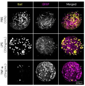
These findings are highly relevant for therapeutic development because neuroinflammation is not a single biological process. Disease states may involve different combinations of microglial activation, astrocytic dysfunction, oxidative stress, and neuronal injury. Models capable of distinguishing between these responses provide a more predictive framework for evaluating therapeutic candidates.
CNS-3D Inflammatory Organoids also support integration of functional calcium imaging with cytokine analysis, immunostaining, cytotoxicity assays, and molecular profiling. This approach addresses another persistent challenge in CNS drug development: many inflammatory assays quantify molecular markers without determining whether those changes correspond to preservation or disruption of neuronal function.
As neurodegenerative drug discovery continues to confront translational issues, interest is growing in models capable of reproducing human-specific cellular interactions and functional neuroimmune responses. Human brain organoid models help bridge the gap between preclinical findings and clinical outcomes by providing a more physiologically relevant framework for evaluating anti-inflammatory therapeutics in the CNS.

To learn more about CNS-3D Inflammatory Organoids, please visit 28bio.com.