![Immunofluorescence image of pituitary cells derived from human pluripotent stem cells. The cells shown here were stained for ACTH (red) and DNA/nucleus (blue) 30 days after differentiation. Similar cells were used in transplantation studies to partly rescue a rat model of hypothyroidism. [Bastian Zimmer, Sloan Kettering Institute]](https://genengnews.com/wp-content/uploads/2018/08/Jun15_2016__BastianZimmer_hPSCDerivedPituitaryCells1334038771-1.jpg)
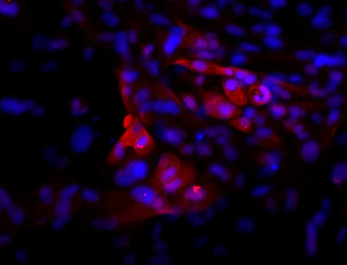
Immunofluorescence image of pituitary cells derived from human pluripotent stem cells. The cells shown here were stained for ACTH (red) and DNA/nucleus (blue) 30 days after differentiation. Similar cells were used in transplantation studies to partly rescue a rat model of hypothyroidism. [Bastian Zimmer, Sloan Kettering Institute]
Signaling progress toward cell replacement therapy for hypopituitarism, scientists based at the Sloan Kettering Institute for Cancer Research have shown that lab-grown pituitary cells can function in rats. The cells, derived from human stem cells, were deployed in a rat model of hypopituitarism, where they secreted hormones important for the stress response as well as for growth and reproduction.
At present, few therapeutic options are available for hypopituitarism, which is linked to dwarfism and premature aging in humans. Moreover, the existing options, such as hormone replacement therapies, are suboptimal. They are expensive, they must be maintained throughout life, and they poorly mimic the body's complex patterns of hormone secretion, which fluctuates with circadian rhythms and responds to feedback from other organs.
By contrast, cell replacement therapies hold promise for permanently restoring natural patterns of hormone secretion while avoiding the need for costly, lifelong treatments. “Cell replacement could offer a more permanent therapeutic option with pluripotent stem cell-derived, hormone-producing cells that functionally integrate and respond to positive and negative feedback from the body,” explained Sloan Kettering’s Bastian Zimmer, Ph.D. “Achieving such a long-term goal may lead to a potential cure, not only a treatment, for those patients.”
Dr. Zimmer led a study that explored a relatively simple way to derive hormone-producing cells. The results of this study appeared June 14 in the journal Stem Cell Reports, in an article entitled, “Derivation of Diverse Hormone-Releasing Pituitary Cells from Human Pluripotent Stem Cells.” The article describes how Dr. Zimmer’s team eschewed a new, but complicated, approach, the generation of 3D organoids, and instead relied on good old monolayer cultures.
“Previous studies have derived pituitary lineages from mouse and human ESCs [embryonic stem cells] using 3D organoid cultures that mimic the complex events underlying pituitary gland development in vivo,” wrote the article’s authors. “Instead of relying on unknown cellular signals, we present a simple and efficient strategy to derive human pituitary lineages from hPSCs [human pluripotent stem cells] using monolayer culture conditions suitable for cell manufacturing.”
Instead of mimicking the complex 3D organization of the developing pituitary gland, Dr. Zimmer’s team exposed hPSCs to a few precisely timed cellular signals, specifically, proteins known to play an important role during embryonic development. Exposure to these proteins triggered the stem cells to turn into different types of functional pituitary cells that released hormones important for bone and tissue growth (i.e., growth hormone), the stress response (i.e., adrenocorticotropic hormone), and reproductive functions (i.e., prolactin, follicle-stimulating hormone, and luteinizing hormone).
These stem cell–derived cells released different amounts of hormone in response to known feedback signals generated by other organs in the body. “hPSC-derived pituitary cells,” noted the article’s authors, “show basal and stimulus-induced hormone release in vitro and engraftment and hormone release in vivo after transplantation into a murine model of hypopituitarism.”
To test the therapeutic potential of this approach, the researchers transplanted the stem cell–derived pituitary cells under the skin of rats whose pituitary gland had been surgically removed. The cell grafts not only secreted adrenocorticotropic hormone, prolactin, and follicle-stimulating hormone, but they also triggered appropriate hormonal responses in the kidneys.
The researchers were also able to control the relative composition of different hormonal cell types simply by exposing hPSCs to different ratios of two proteins—fibroblast growth factor 8 and bone morphogenetic protein 2. This finding suggests their approach could be tailored to generate specific cell types for patients with different types of hypopituitarism. “For the broad application of stem cell-derived pituitary cells in the future, cell replacement therapy may need to be customized to the specific needs of a given patient population,” Dr. Zimmer stated.
In future studies, the researchers plan to improve the protocol further to generate pure populations of various hormone-releasing cell types, enabling the production of grafts that are tailored to the needs of individual patients. They will also test this approach on more clinically relevant animal models that have pituitary damage caused by radiation therapy and receive grafts in or near the pituitary gland rather than under the skin. This research could have important implications for cancer survivors, given that hypopituitarism is one of the main causes of poor quality of life after brain radiation therapy.
“Our findings represent a first step in treating hypopituitarism, but that does not mean the disease will be cured permanently within the near future,” Dr. Zimmer concluded. “However, our work illustrates the promise of hPSCs as it presents a direct path toward realizing the promise of regenerative medicine for certain hormonal disorders.”


