
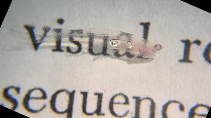
Whitehead Institute researchers have created the first comprehensive whole-body single-cell atlas of an adult vertebrate, the tiny, transparent teleost fish Danionella cerebrum.
The team profiled more than 600,000 cells from across the fish to build the whole-body single-cell transcriptome atlas of which genes are active in which cell. A cell’s transcriptome is the full set of RNA molecules it has made from its DNA at a given time, providing a snapshot of which genes the cell is using.
Together, the data provide a molecular reference for the cell types found throughout Danionella, the patterns of gene activity that distinguish them, and where many of them are located. Offering a new resource for studying brain regeneration, cellular diversity, and investigating how different tissues function together, the atlas gives researchers a baseline for exploring how injury, aging, drugs, or other changes affect cells across the whole animal.
The team has made the atlas publicly available through DigiFish, an interactive resource for exploring cell types and gene activity throughout the animal. Paired with Danionella’s transparency and growing experimental toolkit, the atlas can help researchers identify specific cells and genes to study or manipulate and then follow their activity in the animal.
“Once you have a spatially resolved atlas, you know what cell types exist, but you also know the neighbors of those cell types in this context,” said Kutay Deniz Atabay, PhD, who led the work as a postdoctoral researcher in the lab of Whitehead Institute Member Peter Reddien, PhD. “This gives you access at multiple different levels of complexity, from molecular to cellular to systems.”
Atabay is first author, and Reddien senior and corresponding author of the researchers’ published paper in Science, titled “Whole-body single-cell atlas of an adult vertebrate, Danionella cerebrum,” in which they stated, “This whole-vertebrate transcriptome atlas yields a comprehensive resource for myriad questions in biology and neuroscience.”
Danionella cerebrum is one of the world’s smallest vertebrates (∼12 mm in length), and has the smallest vertebrate brain on record, containing ∼650,000 neurons within 0.6 mm3, the authors wrote. This is approximately four times the size of the Drosophila brain, but with all the major vertebrate brain regions. Viewed from above, Danionella cerebrum offers a rare sight in a vertebrate, in that its brain is visible through the top of its head. “Thanks to its optical transparency, D. cerebrum is arguably unparalleled for noninvasive live whole-brain and whole-body imaging at single-cell resolution in an adult vertebrate …” the team noted.
Since the species was adopted for neuroscience research in 2018 and formally described in 2021, the tiny, transparent fish has drawn rapidly growing interest for its unique biology.
Whitehead Institute researchers recognized that the tiny fish brought whole-animal single-cell profiling within reach, allowing the team to create the first comprehensive map of cell types and gene activity across an adult vertebrate. That perspective could help reveal how cells in different tissues coordinate their responses—and how changes in one part of the body influence others.
Cell atlases have become increasingly detailed, but extending them across a whole adult vertebrate has been difficult for the basic reason of size. Profiling the cellular diversity of larger animals comprehensively is technically demanding and expensive. “The molecular mapping of cell transcriptomes across the entire animal anatomy of an adult vertebrate has not been achieved, largely because most vertebrates are too large for comprehensive molecular access to their cellular and molecular complexity, the investigators pointed out. Danionella—at barely one centimeter long—makes the experiment possible, while its close relationship to zebrafish allows researchers to draw on genetic tools developed for one of biology’s most widely used model organisms. “We reasoned that the extreme miniaturization of Danionella could enable organism-wide cell type profiling and facilitate high-throughput molecular studies,” they continued. “D. cerebrum is also closely related to zebrafish, which allows for the transfer and use of an expansive molecular toolkit, and its genome, similar to many other teleosts, is approximately half the size of the zebrafish genome.“
Atabay first became interested in Danionella in early 2019, while searching for a small, transparent animal in which he could watch the nervous system regenerate. Its miniature size and accessible brain made it especially promising for that work, and the researchers found that the fish has broad regenerative capacity, including in the brain and fins. Reddien’s lab, meanwhile, had already seen the value of a comprehensive whole-animal cell atlas through work on planarians, regenerative flatworms whose small size had made such an atlas possible. Together, those interests led to a larger question.
“Deniz and I had the thought: Would it be possible to get a cell-type transcriptome atlas for a complete vertebrate?” said Reddien, who is also a professor of biology at Massachusetts Institute of Technology and an Investigator with the Howard Hughes Medical Institute. “Here’s an animal where we’d have a chance.”
To build the atlas, the researchers used single-cell RNA sequencing to measure gene activity in individual cells collected from across the brain, spinal cord, eyes, internal organs, fins, head, and trunk. They paired those data with spatial methods that mapped cell types and patterns of gene activity back to their locations in tissues. In their paper the team explained, “We combined regionally stratified single-cell RNA sequencing (scRNA-seq) with whole-animal spatial transcriptomics [multiplexed high-throughput fluorescent in situ hybridization (MERFISH) and hybridization chain reaction fluorescent in situ hybridization (HCR-FISH)] to build, validate, and explore a body-wide single-cell transcriptome atlas.”
The resulting atlas was extensive enough to capture even exceptionally rare cell populations. Looking across the animal revealed patterns that can be difficult to see when tissues are studied separately. In the nervous system, neurons from the same region often had more similar patterns of gene activity, even when they used different neurotransmitters—the chemicals neurons use to communicate—than when compared with neurons using the same neurotransmitter in different regions. Similar patterns have been observed in other vertebrates, including mammals, suggesting that regional and developmental identity may be an important organizing feature of vertebrate nervous systems.
The atlas also revealed signs that adult cells retain a molecular memory of where they belong in the body. The researchers found that fibroblasts, a type of connective tissue cell, maintained region-specific activity of genes best known for establishing different parts of the body during development. Cells in the pectoral and pelvic fins, for example, retained gene programs associated with forelimb and hindlimb development in other vertebrates. What these persistent signals do in adult Danionella remains unknown, but they may help tissues maintain or rebuild the appropriate structures. In planarians, positional information provides molecular guidance that helps regenerating tissues rebuild the correct structures.
The team also used the atlas as a reference for studying Danionella’s remarkable regenerative abilities. After injuring the telencephalon, a region of the forebrain, the missing neurons regenerated and the researchers were able to trace a progression from an early wound and immune response to neural stem-cell activity and the production and maturation of new neurons. “… profiling telencephalon regeneration revealed a temporally ordered molecular cascade, involving signatures of wound response and immune cell recruitment, radial glia proliferation, neurogenesis, and neuronal maturation …” they stated.
For Reddien, the advance presents numerous, diverse opportunities for new science. “There’s been this era of genome sequencing, where you get the complete list of all the genes that it takes to make an organism,” he says. “The transcriptomes of cells tell you how that genome is being utilized in the different cell types. To have the transcriptome of every cell type seems like a big turning point in how one would investigate the biology of an organism.”
In their paper the authors concluded “These findings and resources position D. cerebrum as a powerful model for systems neuroscience, stem cell biology and regeneration, biomechanics and collective behavior, and evolutionary biology.”