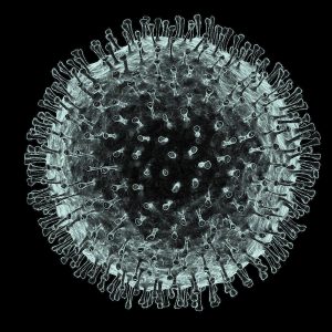
An international team of clinicians and researchers has had an unprecedented opportunity to examine what is likely to represent the early pathology of SARS-CoV-2 coronavirus in humans before symptoms develop. In an article, to be published in the Journal of Thoracic Oncology, a pre-proof of which is now available, Shu-Yuan Xiao, MD, from the University of Chicago Medicine, and a group of clinicians from the Zhongnan Hospital of Wuhan University, describe their examination of surgically removed lung tissue from two patients who had undergone lung lobectomies for adenocarcinoma, but were retrospectively found to have had COVID-19 at the time of surgery.

“This is the first study to describe the pathology of disease caused by SARS-CoV-2, or COVID-19 pneumonia, since no autopsy or biopsies had been performed thus far,” senior author Xiao said. “Since both patients did not exhibit symptoms of pneumonia at the time of surgery, these changes likely represent an early phase of the lung pathology of COVID-19 pneumonia … This would be the only descriptions of early phase pathology of the disease due to this rare coincidence. There would be no other circumstance that this will happen. Autopsies will only show late or end stage changes of the disease.” The team’s paper is titled, “Pulmonary pathology of early phase 2019 novel coronavirus (COVID-19) pneumonia in two patients with lung cancer.”
Although there have been several studies describing clinical features of COVID-19 and characteristic radiographic findings (mainly chest CT scans) no pathologic studies have been conducted based on autopsies or biopsies, the authors noted. “Some of the reasons for the lack of autopsies and biopsies include the suddenness of the outbreak, the vast patient volume in hospitals, shortage of healthcare personnel, and the high rate of transmission, which makes invasive diagnostic procedures less of a clinical priority.”
The researchers were given an unexpected opportunity to examine what is most likely the early lung pathology of COVID-19 when, “fortunately and unfortunately,” they write, they encountered two patients who underwent surgery for lung cancer and were later found to have been infected with SARS-CoV-2 at the time the operations were carried out. As the team explained, “The surgical specimens overlapped in time with the infection, which offered us the necessary specimens to examine the histopathology of COVID-19 pneumonia … To our knowledge, the pathologic findings reported here represent the first for SARS-CoV-2 pneumonia, or 2019 coronavirus infection disease (COVID-19).”
As described in the published paper, the first case was a female patient aged 84 years who was admitted for treatment evaluation of a tumor in the right middle lobe of the lung, which had been discovered on chest CT scan at an outside hospital. She had a past medical history of hypertension for 30 years, as well as type 2 diabetes. Although she was given comprehensive treatment, assisted oxygenation, and other supportive care, the patient’s condition deteriorated, and she died. Subsequent clinical information confirmed that she had been exposed to another patient in the same room who was subsequently found to be infected with the SARS-CoV-2.
The second case was a male patient of 73 years of age, who presented for elective surgery for lung cancer, in the form of a small in the right lower lobe of the lung. He had a past medical history of hypertension for 20 years, which had been adequately managed. Nine days after lung surgery, he developed a fever with dry cough, chest tightness, and muscle pain. A nucleic acid test for SARS-CoV-2 came back as positive. He gradually recovered and was discharged after twenty days of treatment in the infectious disease unit.
Pathologic examinations revealed that, apart from the tumors, the lungs of both patients exhibited edema, proteinaceous exudate, focal reactive hyperplasia of pneumocytes with patchy inflammatory cellular infiltration, and multinucleated giant cells. Fibroblastic plugs were noted in airspaces. The presence of early lung lesions days before the patients developed symptoms corresponds to the long incubation period — generally 3–14 days — of COVID-19.
Although the female patient was never febrile, her CBC profile, especially from post-op day 1, showed “high WBC counts and lymphocytopenia, which is consistent with COVID-19,” the authors stated. “This may be a good clue for early diagnosis in the future.” Case 2 developed a fever a few days after the CT findings, suggesting a delay in symptom development in these patients.” The investigators point out that the time for the early lung lesions or COVID-19 to become severe enough to cause clinical symptoms is “rather long.” Even among patients who do present with fever, commonly used pharyngeal swab PCR tests may still be negative, they suggested, due to the lack of virus in the upper respiratory tract, and despite the presence of pneumonia.
The authors believe that the two incidences they report also typify what commonly occurred during the earlier phase of the SARS-CoV-2 outbreak, when a significant number of healthcare providers became infected in hospitals in Wuhan, and patients in the same room were cross-infected. During this period prevention of transmission was difficult, as many healthcare workers in Wuhan became infected as they were tending to patients without sufficient protection, Xiao noted. Just this week it has been reported that 3,387 health workers in China have been infected with COVID-19, more than 90% of whom in the stricken Hubei province. More than 15 doctors in Wuhan, including young, healthy individuals, have now died of COVID-19 infections contracted while they were taking care of patients.
“The two cases reported here represent ‘accidental’ sampling of the COVID-19, in which surgeries were performed for tumors in the lungs at a time when the superimposed infections were not recognized,” the researchers stated. “These provided the first opportunities for studying the pathology of COVID-19.” Xiao added, “We believe it is imperative to report the findings of routine histopathology for better understanding of the mechanism by which the SARS-CoV-2 causes lung injury in the unfortunate tens and thousands of patients in Wuhan and worldwide.” Further studies are ongoing by the team and collaborators to assess COVID-19 pathology through post-mortem biopsies, and their findings should provide new information on the late changes of this disease.
“It would be beneficial if RT-PCR and/or immunohistochemical stains could be performed on these two cases to further confirm the presence of the viruses that may be associated with the pneumonia,” the investigators further commented. “Unfortunately, these tests are currently under development, and adaptation to tissue specimens is not yet available. Nevertheless, we believe it is imperative to report the findings of routine histopathology for better understanding of the mechanism by which the SARS-CoV-2 causes lung injury in the unfortunate tens and thousands of patients in Wuhan and worldwide.”
The authors also caution the need for “universal precaution” in surgical pathology laboratories, which should be mindful of the potentially infectious nature of all fresh specimens. In China, most surgical specimens are received already fixed in formalin. However, the investigators stressed, “for larger specimens the center of a specimen may not be sufficiently fixed and still pose potential risk for infection. Therefore, proper PPE with surgical masks or N95 respirators are worn all the time in the gross room. Fortunately thus far to our knowledge, no cases of pathologists being infected by COVID-19 had occurred.”


