
Quantitative spatial biology is coming of age. The desire to learn beyond the surface has always been present but now the ability to deepen that knowledge with real data is catching up. High-plex spatial proteomics can provide details into single-cell molecular measurements within a tissue context. This advanced quantitation moves imaging forward to enable reproducibility, mechanistic insight, biomarker validation, patient stratification, and regulatory-grade evidence.
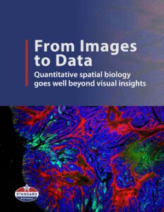
Tissues can be seen as structured ecosystems in which cell identity, function and interactions depend on precise spatial organization and local microenvironments. Adding quantitative metrics offers the opportunity to not only directly investigate changes in cell behavior and tissue heterogeneity but also the ability to compare these patterns across samples, disease states, individuals, or treatment conditions.
When investigating cancer progression, immune response to disease or therapeutic response, visualizing and quantifying the spatial organization of proteins across a tissue enables a look into patterns of cell activity as well as advanced modeling and predictive analytics that bring us ever closer to targeted therapeutics and personalized medicine. This eBook introduces the latest trends in quantitative spatial biology and how spatial proteomics is accelerating their adoption and success.