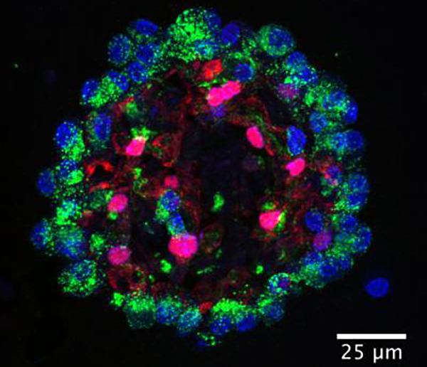
A pair of studies have delved into lung cell development, or rather re-development, uncovering clues to how lung tissue maintains itself or recovers from injury and disease. Both studies focused on mesenchymal cells, which are generally thought to play a supportive role in maintaining lung structure. These cells, it turns out, help manage the regeneration of lung tissue. They keep lung cell subpopulations in proper balance—or not, if, for example, they generate an excess of scar tissue while failing to produce sufficient numbers of functional cells.
One of the studies stratified lung mesenchymal subsets by using single-cell and population-based RNA sequencing. These techniques, deployed by scientists based at the University of Pennsylvania School of Medicine, made it possible to identify five distinct cell types in the mouse lung. Focusing on two of the five cell types, the scientists found that “each mesenchymal lineage has a distinct spatial address and transcriptional profile leading to unique niche regulatory functions.”
Details of this work, which helped the scientists produce a spatial and transcriptional map of the lung mesenchyme, appeared September 7 in the journal Cell, in an article entitled, “Distinct Mesenchymal Lineages and Niches Promote Epithelial Self-Renewal and Myofibrogenesis in the Lung.” In this article, the University of Pennsylvania team described how it analyzed secreted molecules and surface cell receptors from the cells of interest, and compared this information to databases of known secreted molecules and receptors on adjacent cells.
“The mesenchymal alveolar niche cell is WNT responsive, expresses Pdgfrα, and is critical for alveolar epithelial cell growth and self-renewal,” the article’s authors detailed. “In contrast, the Axin2+ myofibrogenic progenitor cell preferentially generates pathologically deleterious myofibroblasts after injury. Analysis of the secretome and receptome of the alveolar niche reveals functional pathways that mediate growth and self-renewal of alveolar type 2 progenitor cells, including IL-6/Stat3, Bmp, and Fgf signaling.”
The “good” mesenchymal alveolar niche cells (MANCs) are found in niches or compartments near the alveoli to promote renewal of gas-exchange cells. They may play a key role in maintaining the alveoli during the normal life span of the adult. Dysfunction or loss of MANCs may contribute to diseases such as COPD, which involves loss of alveoli and decreased lung function. The role of the “bad” Axin2+ myofibrogenic progenitor cells (AMPs) is to form scar tissue during wound healing. However, AMPs may grow out of control, potentially leading to diseases such as IPF.
“One of the most important functions of these cells is to balance the repair and regeneration response after injury which occurs often due to the lung's continual assault from the outside environment,” said Edward E. Morrisey, Ph.D., senior author of the study and a professor of cell and developmental biology and director of the Penn Center for Pulmonary Biology.
Next, the researchers aim to identify these cell types in humans. Dr. Morrisey indicated that the Penn team wants to target MANCs for promoting regeneration while inhibiting AMPs to reduce the fibrotic response after injury. Knowing the detailed molecular differences between these two cell types should help in the next generation of targeted therapies, such as nanomedicine.
The other study that investigated mesenchymal lineages used genetic lineage tracing, single-cell RNA sequencing, and organoid culture approaches. This study, which was conducted by scientists based at Boston Children’s Hospital, focused on distinct mesenchymal niches that drive airway and alveolar differentiation. The study’s authors expressed the hope that their work could help in developing new treatments for such lung disorders as asthma and emphysema, by revealing ways to manipulate natural systems for treatment purposes.
This study (“Anatomically and Functionally Distinct Lung Mesenchymal Populations Marked by Lgr5 and Lgr6”) also appeared September 5 in the journal Cell. The study identified region- and lineage-specific crosstalk between epithelium and their neighboring mesenchymal partners. These findings, the study’s authors asserted, may provide a new understanding of how different cell types are maintained in the adult lung.
In particular, the study highlighted that Lgr5 and Lgr6, well-known markers of stem cells in epithelial tissues, are markers of mesenchymal cells in the adult lung. “Lgr6+ cells comprise a subpopulation of smooth muscle cells surrounding airway epithelia and promote airway differentiation of epithelial progenitors via WNT-Fgf10 cooperation,” the study indicated. “Genetic ablation of Lgr6+ cells impairs airway injury repair in vivo. Distinct Lgr5+ cells are located in alveolar compartments and are sufficient to promote alveolar differentiation of epithelial progenitors through WNT activation.”
“Modulating WNT activity,” the article’s authors added, “altered differentiation outcomes specified by mesenchymal cells.”
“All of these cell types are interacting with each other to repair and regenerate lung tissue,” explained Carla Kim, Ph.D., the study’s senior author and a researcher at the Stem Cell Research Program at Boston Children's Hospital. “What we learned is that mesenchymal cells are telling the epithelial stem cells what type of lung cell they should make.”
“If you have a disease like emphysema or bronchopulmonary dysplasia, where alveolar cells are not being repaired, stimulating the WNT pathway could be important,” Dr. Kim continued. “When WNT is blocked, you get the alternate kind of cell. But if you add WNT back in, it goes back to usual.”
For airway problems like asthma, blocking WNT or finding a way to boost the formation of LGR6-bearing cells could be approaches to explore to increase production of airway cells. “We found that if you injure mouse lungs so airway cells are killed off, you need LGR6+ cells to repair that kind of damage,” noted Dr. Kim. She acknowledged, however, that there's much more to be learned: “What we really want to know next is, what are all the secreted factors these cells make that can control cell fate? Those could be potential therapeutic targets.”


