- Time:
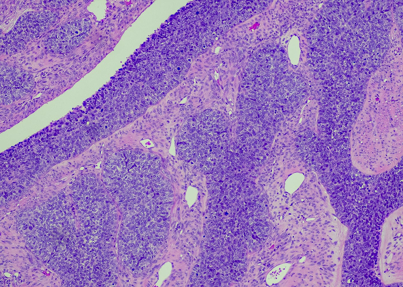
Scientists gain a deeper understanding of different cancers when they analyze a range of omics data types. New tools like multiplexed imaging provide functionality for studying omics data in its tissue environment, shedding light on the complex pathways, networks, and mechanisms that cancer cells use to communicate and survive. By imaging RNA and protein data in the same tissue section, scientists can uncover meaningful insights about the spatial landscapes and crosstalk signaling networks of cancer cells, and further understand their role in modulating therapeutic outcomes.In this GEN webinar, Dr. Sammy Ferri-Borgogno, an instructor in the department of gynecologic oncology and reproductive medicine at MD Anderson Cancer Center, will discuss how a multi-omic approach to tissue imaging can reliably characterize the tumor microenvironment in ovarian cancer. She will discuss the capabilities of Imaging Mass Cytometry, ™ which is used to generate high-dimensional spatial data from tissues at subcellular resolution, and why this technology is ideal for large studies involving many samples. During the webinar, you’ll learn:
- The importance of spatial cellular organization in the tumor microenvironment and its role in modulating clinical outcomes.
- How to apply a multi-omic analysis approach (proteomics, transcriptomics) to a tissue section to precisely identify novel therapeutic targets and reveal mechanisms of resistance to chemo- and immune- therapies.
- How leveraging higher-plexed Imaging Mass Cytometry™ can characterize spatially resolved mRNA (spatial transcriptomics) and protein (spatial proteomics) simultaneously.
A live Q&A session followed the presentation, offering a chance to pose questions to our expert panelist.

Instructor, Dept. of Gynecologic Oncology and Reproductive Medicine
MD Anderson Cancer Center
Webinar produced with support from:
