
July 1, 2017 (Vol. 37, No. 13)
Possibilities for Basic Research, Drug Discovery, and Therapeutics Expand Geometrically
Once confined to disease modeling and drug safety testing, 3D cell culture is expanding in multiple directions, exploring new possibilities in basic research, drug discovery, and therapeutics. In basic research, 3D cell culture is being used to establish models of disease and unravel the mysteries of differentiation. In drug discovery, 3D cell culture is demonstrating that it isn’t just for toxicology—it can be adapted for target identification and drug candidate screening, too. And in therapeutics, 3D cell culture promises to realize several exciting applications, from patient-specific drug-efficacy screening to the manufacturing of bioprinted organs.
These possibilities are coming within our grasp because 3D cell culture, unlike 2D cell culture, has depth—and not just in the obvious geometrical sense. 3D cell culture captures structural and organizational variations, and hence physiological variations, that can help it represent real, living tissue far better than 2D cell culture ever could. 3D cell culture, besides being more convenient than animal models (and relatively free of ethical issues), permits extraordinary control—from the genetic level upward.
In addition to raising the possibility of patient-specific models, 3D cell culture could progress beyond relatively simple self-aggregated spheroids to replicate all manner of developmental contingencies in environments consisting of multiple cell types and diverse extracellular elements. And if that means manipulating cells magnetically or embedding them in microfluidic structures, so be it.
Realizing these heady possibilities is a daunting challenge. It’s on the order of transitioning from checkers to chess—three-dimensional chess. Fortunately, it isn’t beyond our panel of experts. Consider them the grandmasters of 3D cell culture. For your edification, they have answered our questions about 3D cell culture’s opening game, as well as how the endgame may develop.
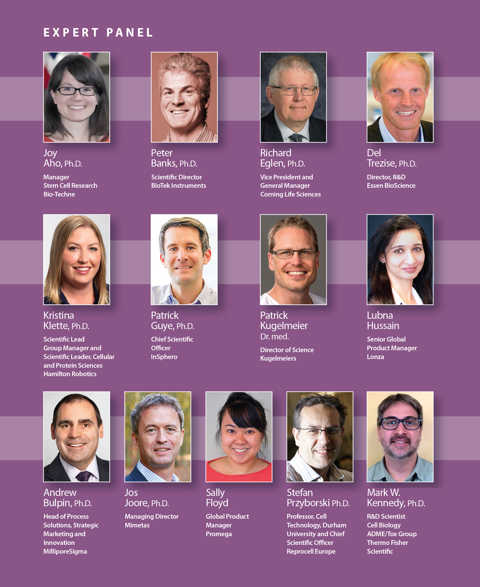
GEN’s Expert Panel
GEN: Are 3D cell culture models as strongly focused as ever on drug safety testing, or are they finding new applications?
Dr. Aho: The focus of 3D cell culture models has definitely expanded beyond drug safety testing. It is becoming increasingly clear that these models mimic cells in vivo at a greater capacity than traditional cell culture.
In addition to drug toxicity, 3D models are progressively being developed and used in developmental biology research, disease modeling, and regenerative medicine. 3D models also provide an enhanced system for drug discovery. Because they better recapitulate disease in vitro, 3D models have the potential to accelerate the testing timeline for drug efficacy studies.
Dr. Banks: Another major application area of 3D cell culture models is in oncology. Spheroids in both media and Matrigel® can be used as surrogate models of tumor proliferation and tumor invasion. Automated brightfield or fluorescence microscopy is typically used for spheroid or invadipodia area measurements. In addition to spheroids, collagen-based scaffolds that encourage cell aggregation into tumoroids have been used for immunotherapy applications such as natural killer cell cytotoxicity assays. Finally, magnetic particles have been used to bioprint cells for cell migration and invasion experiments.
Dr. Eglen: We would argue that 3D cell culture models have been used for many years in basic research and disease modeling, notably in cancer research—this was, after all, one of the original applications of Corning® Matrigel®, a naturally occurring extracellular matrix for us in 3D cell culture. That said, it is true that 3D cell culture models are increasingly being used in preclinical lead optimization, particularly in evaluating potential compound toxicity and metabolic liability.
Furthermore, disease research areas are expanding to include neurology, stem cell research, cell therapy, and (potentially) tissue engineering. Perhaps the most exciting work is the development of 3D technologies for the optimal production of patient-specific cells, either for compound testing or possibly cell therapy.
Interestingly, spheroids derived from stem cells grown in 3D models show improved “stemness,” that is, characteristics that may lead to increased efficacy in regenerative medicine. Researchers have seen that spheroids display enhanced anti-inflammatory, tissue regenerative, and reparative responses, as well as better post-transplant survival of mesenchymal stem cells.
Autologous tissue for transplantation may also come from organoids produced via 3D cell culture. For example, renal organoids derived from pluripotent stem cells have been successfully transplanted under the renal capsules of adult mice. Clearly, research in this area is advancing rapidly, probably due to a convergence of several multidisciplinary fields, ranging from bioengineering, materials science, phenotypic screening, and cell biology.
Dr. Trezise: Drug safety continues to be a significant application area for 3D models. This application area has become only more interesting as more data has become available indicating that 3D models offer translational benefits. In addition, there is a growing trend to develop 3D models that can advance developmental biology, target validation, and drug efficacy studies. This trend is particularly evident in the field of oncology, where researchers are combining patient-specific tumor cells and 3D cell culture methods to create tumor organoids. These mini-tumors are being used to determine sensitivity to combinations of different chemical, biological, and cellular therapeutics in the context of personalized medicine.
Dr. Klette: 3D cell culture models are widely used for drug safety testing, such as studying hepatic injury from compound screens, and for examining drug metabolism using 3D hepatocyte models. In personalized medicine, however, patient-derived primary 3D models are being used for cancer screening in biotherapeutics. Here, 3D models provide enhanced physiological relevance to determine drug efficacy and potential impacts on carcinogenesis, metastasis, and tumor reoccurrence. If we look outside drug discovery and biologics, we notice that areas such as regenerative medicine and cell therapies can take advantage of 3D models as a predictor of disease and (when scaled to therapeutic levels) as a disease treatment.
Dr. Guye: 3D cell models are applied throughout the biomedical and life sciences. 3D technologies that are compatible with high-throughput screening are used not only for screening purposes, but also for target and hit validation, lead optimization, and investigational toxicology.
Basically, given their ability to extend cell lifetimes and incorporate multiple cell types, 3D models are increasingly finding their way into basic research, where they are helping to recapitulate disease progression and assess the impact of certain genes and pathways on disease progression/prevention—activities that help scientists define adverse outcome pathways. Importantly, we expect human 3D cell culture models to significantly reduce the percentage of drugs that progress to clinical trials and fail due to lack of efficacy.
Dr. Kugelmeier: The focus on drug safety testing is still valid, and sophisticated organoid models might contribute to even more accurate drug safety testing because of increased physiological fidelity of these models. But there are also significant new research areas. Combining organoid technology with stem cell biology could lead to therapeutic applications. Also, cancer research—especially cancer research that focuses on cancer stem cells—needs 3D models. Of these models, cell spheroids are among the most important. Sophisticated cell-spheroid platforms not only allow research but also provide drug-testing possibilities using patient cells for personalized medicine. Finally, these platforms may enable therapeutic applications with stem cell spheroids in regenerative medicine.
Mrs. Hussain: The focus for 3D cell culture methods is still the drug safety testing that occurs before in vivo testing. Recently, there has been a renewed interest in phenotypic drug screening to discover new drug targets. With this shift, there is growing emphasis on bridging the gap between phenotypic screens and 3D methods. Phenotypic screens, in vitro, were traditionally carried out using 2D methods that do not take into account the complexity of the in vivo environment. 3D methods are now sought to build biologically relevant models that are more predictive of phenotypic response to new drug targets.
Dr. Bulpin: Applications continue to expand for 3D models, including the development of specific disease models and complex tissue models that can be used for basic research as well as drug discovery. Another promising area for 3D models is personalized medicine. Several types of cells can be used in these models including “immortalized” cells, genetically engineered cells, induced pluripotent stem cell–derived cells, primary human cells, and patient-derived cells (including patient-derived xenografts). Another potential research avenue is engineering 3D tissues for organ transplants.
Dr. Joore: Over the last year, we observed a growing interest in 3D tissue models that could be used in studies of disease processes, whether the studies emphasized screening or efficacy analysis. These are, I think, two sides of the same coin. Once researchers realize they need better predictive models for safety testing, they start to see that improved models would also have potential for discovery and development. Molecule-to-molecule screens have generated lots of very specific inhibitors, but not so many therapies. Researchers are now starting to appreciate the richness of 3D model data, especially in combination with the throughput of our organ-on-a-chip platforms.
Ms. Floyd: Cancer researchers and developmental biologists have certainly benefitted from 3D cell culture models, which are more physiologically relevant than are 2D systems to the study of cellular differentiation. Further, 3D in vitro systems are well positioned to obtain approvals from authorities such as the Organization for Economic Co-operation and Development Organization (OECD). The OECD and other bodies are considering alternatives to whole-animal testing, including alternatives that can accomplish skin-sensitization studies for the safety assessment of chemicals.
Prof. Przyborski: What has changed more recently is the ease of access to innovative technologies on the market that enable researchers to more readily practice 3D cell culture routinely. 3D cell culture has had impact in multiple areas in basic research, drug screening, and safety assessment. Researchers are now looking to 3D technologies to create more sophisticated models that are representative of real human tissues. Investment in more advanced in vitro assays at an early stage will improve predictions of drug action and inform the decision-making process as to whether to further invest in a particular drug candidate.
Dr. Kennedy: 3D cell cultures continue to be extensively explored for drug safety screening; however, there is a growing interest in expanding the use of more complex 3D models into areas such as disease modeling and precision medicine. For example, preclinical hepatic research is now looking to exploit the benefits of spheroid cultures by building 3D co-culture models that consist of multiple primary liver cell types to create new models of hepatic and biliary disease. Likewise, stem cell–derived organoids are opening the possibility of tailoring therapeutic regimens to patients’ genetic makeups and to identify the best treatment options.
GEN: What is the biggest challenge facing drug developers that use 3D cell culture models?
Dr. Aho: Robustness and consistency are major challenges. Individual research groups have used varied 3D cell culturing methods. Consequently, there has been a lack of model standardization within the field of drug development. Current methods have mixed success rates between research groups and between starting samples, leading to long-term concerns over sample consistency and robustness.
Many 3D models rely on cell clusters, the sizes and shapes of which are inherently variable between samples. These inconsistencies can lead to challenges in downstream data analysis methods. Similar challenges may arise from issues of tissue accessibility when cell clustering methods are used.
Dr. Banks: Many 3D cell culture technologies are relatively expensive. Assay costs/well are typically more than $1 and sometimes exceed $10. This precludes their use in early preclinical drug discovery procedures, such as primary screens, where assay costs/well should be << $1.
Dr. Eglen: There are still several challenges that remain in the adoption of 3D cell culture technologies, though none of them appear to be insurmountable. For example, the systematic assessment of compound pharmacology is challenging partly because some 3D cell culture models, such as spheroids or organoids, may exhibit significantly more complex morphology and function than cells simply grown in 2D culture. Indeed, cell populations in 3D culture can be very heterogeneous due to variations in complexity, size, morphology, architecture, and (ultimately) assay protocols—such as protocols for cell-based high-throughput screens.
Only a few high-throughput screening or compound-profiling campaigns have directly compared 3D data to 2D data. Whilst, the field still debates the significance of the differences between 3D culture results and 2D culture results, there is a growing consensus that 3D culture is better than 2D culture at replicating in vivo conditions.
Dr. Trezise: It makes little sense to develop advanced 3D cell culture models with input cells that have inherently low relevance or predictivity. Researchers are therefore having to move away from traditional cell lines to primary cells and stem cell–derived tissue, preferably human. The added complexity and cost in sourcing, handling, and characterizing these cells is certainly challenging. In addition, most assay detection methods are optimized for traditional 2D cell cultures, so drug developers are also having to rethink how best to make meaningful analyses from 3D systems, especially in regard to relevant readouts at the required throughput.
Dr. Klette: Technical and optimization expertise is required in 3D cell culture model development. Proper 3D culture handling is important. It should be easy to use and manipulate 3D models without harming them or the cells they contain. For example, cells extracted from a matrix or scaffold shouldn’t undergo lysis due to applied shear forces. In 3D models of cancer, assessments of drug compound penetration may require gradient geometries to be calculated, and they may be further complicated if heterogeneous cell models are used.
From a production standpoint, no single 3D model suits all needs. Consequently, the challenge remains for drug developers to find what works for them in terms of efficiency, reproducibility, long-term culture maintenance, and scalability.
Dr. Guye: A major challenge is combine ease of use, scalability, reproducibility, and the ability to directly fit 3D cell culture models into existing drug screening pipelines—and to do so at a reasonable price. Another challenge is that even though 3D models are being employed more frequently in drug development, they lack a similar-sized data bank compared to the former standard models. This may lead to discordant results. Therefore, harmonization of model qualification and validation would be highly beneficial, enabling developers to compare different models, make the best use of the most appropriate models, and integrate the resulting data into decision-making processes.
Dr. Kugelmeier: Although cell spheroids offer advantages such as reproducibility, precision, and versatility (they offer many application possibilities), they also pose several challenges. When cell spheroids are used, cluster size directly impacts diffusion distance of every molecule entering every one of the cluster’s cells. Consequently, cluster size needs to be controlled to obtain reliable data. The control of cluster size is achieved almost exclusively by means of hanging drops. But besides lacking medium change capabilities, the generation of hanging drops is very labor intensive, and professional hanging drop platforms are very expensive. And even these platforms lack scalability; that is, they cannot generate “sized” clusters in large numbers. For screening applications and more, easier scalability is needed.
Mrs. Hussain: 3D cell cultures require time and resources for optimization in order to build good in vitro models, particularly for multilayer complex models. Additionally, using 3D cultures with standard cell analysis methods that were developed to support 2D cultures can be challenging. In drug discovery, shortening timelines and increasing throughput is key.
Set-up times required by conventional 3D cultures have exceeded those required by 2D cultures. However, automated workflows have recently been adapted and validated for use in 3D cell culture system studies, enabling numerous replicates to be configured simultaneously using an identical technique for each culture. Additionally, more protocols are being developed by commercial suppliers offering 3D culture tools to ease the adoption for researchers. These factors have helped reduce the initial set-up time required, aiding workflows in high-pressure drug discovery laboratories.
Dr. Bulpin: The biggest challenge for drug developers is determining whether a 3D model is more predictive than current 2D models. The 3D models are certainly more physiologically relevant and reflective of the in vivo environment, but whether they improve predictivity remains to be determined.
A second important challenge is the throughput of 3D models. Many of these new 3D models have low throughput and thus cannot be used as a primary screen in the traditional sense. Additional challenges include connecting various organ models (to simulate whole-body physiological responses), formulating compatible media, and accessing relevant cell types.
Dr. Joore: A recent article in Nature Medicine describes the key issues as throughput, validation, and the ability to start solving pharma’s biological problems. We have addressed these issues by incorporating throughput in the basic design of the OrganoPlate®, a microfluidic, 384-well plate that can support up to 96 tissue models. With this as a starting point, we have worked with pharma to develop models that tackle their problems, from safety to efficacy to the identification of new compounds based on phenotypic screens.
Validation still poses difficulties. For example, validation studies that compare new models with “gold standard” systems may inspire little confidence because the gold standards are seldom very predictive; however, studies that rely on retrospective compound screens may be helpful.
Ms. Floyd: For users of 3D cell culture models, it is a major challenge to choose a physiologically relevant method that is predictive of in vivo conditions, cost effective, and amenable to high-throughput operations. There are few “off the shelf” assays that pertain to 3D models and have validated protocols. In hydrogel systems, diffusion and penetration of reagents can be ineffective. Spheroid penetration can also pose as a challenge when biologics are tested. Highly sophisticated methods, such as organ-on-a-chip methods, may simulate physiological more closely, but they are relatively expensive and resistant to high-throughput operations, compared to methods that make use of ultra-low attachment technology.
Prof. Przyborski: Challenges that may limit the uptake of 3D cell culture technology include accessing the technology (commercial off-the-shelf systems would be ideal); developing custom 3D cell culture assays (clients’ needs should be addressed); acquiring 3D cell culture skills and knowledge (in-house expertise for creating and working with 3D culture models would be advantageous). These challenges can be met by partnering with experts and organizations that specialize in 3D cell culture technologies to build custom solutions to help improve the value and predictive accuracy of in vitro assays using 3D cell culture technology.
Dr. Kennedy: One of the biggest challenges drug developers must face is large-scale screening. Consistently generating necessary volumes of 3D spheroids is neither trivial nor economical, and cytotoxicity screens often parse thousands of compounds. Moreover, organoids and microphysiological systems require complex growth media and specialized reagents that impact the ability to generate large-scale cultures. At Thermo Fisher Scientific, we are leveraging our expertise in cell culture reagents to address these problems. We believe that understanding these issues will go a long way toward defining the best application of 3D cell models in the drug discovery and development process.
GEN: Is there technology on the horizon that will significantly impact 3D cell culture models?
Dr. Aho: We believe that new tissue model systems will address many current pain points of 3D cell culture. One such system is based on technology developed by the Multiclonal Therapeutics research team of Wa Xian, Ph.D., and Frank McKeon, Ph.D. It allows for the derivation and expansion of ground-state stem cells from adult tissue.
These cells can then be differentiated back into the host tissue, which then maintains its native architecture, regional specificity, and (if isolated from diseased tissue) disease phenotype. This system is robust across samples, provides better 3D model standardization and accessibility, and removes the structural inconsistencies seen in cell clustering methods.
Dr. Banks: Recently, we have been working with a 3D magnetic bioprinting technology for applications such as migration and proliferation. We have also successfully used the technology for complete “walkaway” automation of mesenchymal stem cell differentiation into chondrocytes. During the two-week differentiation period, chondrocyte function in 3D models was far superior to that in typical microplate models.
Dr. Eglen: In a rapidly evolving field such as 3D cell culture, technologies continue to evolve that provide increasingly easier adoption to 3D models, and simpler, more robust protocols. These technologies include microtiter plates for culturing and high-throughput screening assays of spheroids; natural and synthetic hydrogels for the development of extracellular matrices upon which a variety of cells can be grown; and permeable supports that facilitate complex cell culture models including multilayered tissues, migration/invasion assays, and co-culture applications.
Further out on the horizon may be applications such as organs-on-chips, hydrostatic flow technologies, microfluidics, and 3D bioprinting. To the latter point, printing an intact organ remains elusive, but 3D-bioprinted bladders, tracheal grafts, bone, and cartilage have proven to be functional. Potentially, these models will allow observations of cellular responsiveness to drug candidates in real time; that is, 3D culture may move to 4D culture!
Dr. Trezise: Improvements in the 3D cell culture toolkit include cell-imaging modalities, cell biomatrices, and stem-cell harvesting and differentiation protocols. In addition, 3D cell cultures are incorporating continuous live-cell analysis and imaging, raising hopes that researchers will gain the ability to follow the development, maturation, and functional properties of 3D cell cultures over time, and to do so without resorting to methods that introduce perturbing influences. Only when 3D cell cultures can be created, manipulated, and analyzed as readily as can 2D cell cultures, will the true promise of 3D biology be realized.
Dr. Klette: New spheroid creation technologies continue to expand the field. For example, one of our partner companies uses a patented micromolded microplate to promote self-aggregation in cell cluster production. But no matter how the 3D spheroids are formed, automation is a critical workflow component. Automation increases throughput, streamlines workflows, enhances reproducibility, and decreases variability compared to manual methods.
Additionally, reagent and material costs can be reduced when miniaturizing and automating 3D methods, while minimal hands-on time frees users to focus on other laboratory tasks. Finally, automation monitors and tracks the entire process from research and development, through scale-up, and on to manufacturing.
Dr. Guye: A trend to monitor is the application of genetically engineered tissues and reporter cell lines, as well as tissues derived from human induced pluripotent stem cells. Although these models are promising, they are, in many cases, incapable of delivering adequate performance.
From the technology perspective, there are already many methods and technologies available to make 3D cell culture models to cover most organs in the body, and many of these models are tailored for use in distinct phases of drug development. The current technology, however, will not allow developers to take full advantage of 3D models until it incorporates certain advances, such as microphysiological systems capable of interconnecting 3D models, more thorough pharmacological data, and implementation of clinically relevant biomarkers into assays adapted for use with advanced models.
Dr. Kugelmeier: Responding as the representative of a company that is developing a novel 3D cell culture technology, my answer is biased. But I am not alone in thinking that basic science has never been so close to realizing so many clinical applications. The current state of scientific and technological developments, especially with stem cells, inspires bold visions about the future of medicine.
Helping to realize the new clinical applications are several “workhorse” technologies. One of them, 3D cell culture, is effectively being harnessed now that the scalable production of sized, stem cell–quality clusters is possible. Other workhorses include gene editing, extracellular matrix, single-cell-handling, and imaging technologies.
Dr. Bulpin: Genome-editing tools (such as CRISPR), 3D bioprinting, microfluidics, and imaging technologies are all advancing rapidly and will likely have a significant impact on 3D cell culture in the near future.
Dr. Joore: I personally believe that organ-on-a-chip technology in combination with induced pluripotent stem cells, organoids, and CRISPR/Cas9 presents a tantalizing opportunity to generate complex biology, and cells carrying any genetic background may be used. Think about complex, intractable diseases such as central nervous system disease, neurodegenerative diseases, and cancer. Machine learning and artificial intelligence will be essential to analyzing the multifactorial output of such 3D, multicell-type models. And finally, we’re working on novel ways to create even more physiologically relevant models in the OrganoPlates® by adding more tissue lanes, functionalities, and readouts.
Ms. Floyd: The development of new instruments that provide real-time imaging and plate-reading capabilities, such as the Essen Bioscience IncuCyte® Live Cell System, will be largely enabling for 3D culture systems. The ability to detect cell-specific markers in mixed-cell populations will be particularly useful with 3D models. Finally, 3D bioprinting will drive research and discovery in new and exciting ways, allowing the creation of tissue and organs that will provide more biologically predictive models.
Prof. Przyborski: 3D cell culture is a tool that enables researchers to improve the growth and function of cells. It will play an important role in the creation of more sophisticated tissue models that more accurately mimic tissue structure. 3D culture combined with other technologies, such as oxygen control and fluid flow, will enhance such models still further as surrogates for real tissue. These technologies in combination with human stem cell science will open new opportunities where renewable sources of cells can be generated to create robust and reproducible 3D models of human tissues for use in drug development.
Dr. Kennedy: Stem cell–derived organoids are composed of multiple cell types. Although their size and differentiation time can be controlled, the exact cellular composition may vary significantly, thus making data interpretation difficult and inconsistent. One possible solution is the adoption of “designer matrices” to guide the differentiation process. These are synthetic and tunable extracellular matrix proxies that can be designed to more accurately mimic the physical properties and chemical compositions of native extracellular matrices. In turn, designer matrices may lead to more consistent and mature organoid cultures that improve the quality of the data being generated.
3D Phenotypic Assays for Personalized Medicine
Drug discovery today is mostly based on parameter research, i.e., to measure individual factors like gene-expression levels, protein levels, protein activity, and localization, and then try to extrapolate this into in vivo function for treatment in humans.
The extrapolation of the effect in a 2D cell culture to the in vivo human situation has often resulted in less favorable downstream effects of limited efficacy or toxicological side effects. One example is the screening of anticancer drugs performed in oxygen-rich 2D cell culture in which high-specific metabolism gives very different chemical hits compared to the screening on hypoxic 3D cell cultures. The oxygen-starved situation will be a closer match to the situation in solid tumors in vivo. Performing a metabolism-based screening with phenotype-based readout provides the possibility to screen closer to the real-life situation.
Technology like the calScreener calorimetry-based assay provides a direct measurement of the cell activity without labels or additions of any kind, according to Magnus Jansson, CSO, Symcel, who adds that it is not measured via a proxy measurement; it is a direct measurement of the energy release.
“Direct metabolic readout (DMR) provided by the technology gives the scientist access to an unbiased assay where the effects of treatment are studied in a correct biological context resulting in greater predictive value for cost-effective and rapid drug development,” says Jansson. “The technology can be used to bridge the gap between screening isolated components to 2D cell cultures to 3D tissue to the in vivo human situation, thus increasing the predictive power of the scientific hypothesis in drug development.”
The extension of 3D-based drug discovery would be to apply the same assay principle on actual patient tissue samples to evaluate the best possible treatment based on the real patient response in phenotype response based and personalized medicine, continues Jansson, who predicts a resurgence of calorimetry cell-based assays in drug discovery and personalized medicine.

