December 1, 2017 (Vol. 37, No. 21)
Beyond Immune Cell Characterization
The immune system is continually surveilling the body—looking for pathogens, xenobiotics, and other non-self signals. The various cell types which constitute the immune system are, thus, incredibly dynamic and capable of upregulating the processes that go into handling these insults on a time scale of minutes to hours. Many cell types in this system are capable of activation to secrete cytokines, rapidly proliferate, or otherwise communicate to surrounding cells that there is a pathogen to consider. Upon clearance of the pathogen, the cell population must then contract in a controlled manner. Furthermore, in some cell populations (e.g., T cells) a subset of the cells are retained as long-lived memory cells to protect and prime the system for future insults. To enable this dynamic function, immune cells integrate internal cellular signals, cues from the tissue microenvironment, and cellular metabolic activity.
The dynamic nature of immune-cell function poses a unique challenge to the analysis of these cell types. Many of the typical methods that are used to quantify immune-cell function are in fact not “functional” assays at all. Typically, researchers utilize snapshot or point-in-time methods to interrogate whether the cells are activated. Alternatively, many scientists screen for marker expression to characterize which cell subpopulations are present. For example, expression of the CD69 surface antigen is frequently used as a marker of T-cell activation. Notably, this approach is most relevant for defining or quantifying a subpopulation of cells used in an experiment. Surface markers used to identify activated cells often require many hours to become present on the cell surface. While these antigens are often used to inform experiments, they don’t provide the temporal discrimination to see activation kinetics or allow researchers to modulate immune functionality in real time.
Recent advances in the field of immunology identify cell metabolism as a critical regulator of immune-cell function. Indeed, changes in cellular metabolism are not only permissive for altered cell function but are, in fact, sufficient to cause these changes. Metabolic reprogramming occurs in the order of minutes to allow changes in cell function, providing a novel and unique marker for functional analysis. Agilent Seahorse XF technology is at the forefront of the field of immunometabolism, enabling researchers to measure cellular metabolic activity acutely and continuously, as cells are stimulated in a real-time manner. Label-free sensor technology coupled with integrated drug/compound injection ports and real-time interrogation provide a powerful platform to quantify and modulate the dynamic function of immune cells in a manner that is complementary to traditional point-in-time analysis methods.
To illustrate the principles described above, we present three vignettes of immune-cell activation, each of which highlight a unique aspect or need for researchers attempting to uncover the points of regulation in their cell models.
Access to the Earliest Events in T-Cell Activation
Recent data from the lab of Christoph Hess, M.D., at the University of Basel, Switzerland, embraces the idea that T-cell activation is correlated with rapid shifts in cellular metabolism.1 This shift in metabolism is especially prominent for the glycolytic pathway, which provides the necessary cellular building blocks and energy required for high rates of cellular proliferation. Using XF technology to noninvasively measure glycolytic rate, activation of T cells after stimulation with anti-CD3/anti-CD28 beads is evident within minutes (Figure 1). This rapid increase in glycolytic rate has been observed in both CD4+ and CD8+ T-cell subsets.
Conventionally, the progression of T-cell activation is measured in terms of changes in cell size/morphology, interleukin/interferon expression, and/or lactate efflux. The increase in glycolytic rate observed in this study is directly correlated with these approaches. Importantly, modulating T-cell activation by adding inhibitors of the glycolytic pathway also results in decreased activation when measured using alternative approaches.

Figure 1. Rapid detection of T cell activation using Agilent XF technology. Human naïve CD4+ T cells were measured using an Agilent Seahorse XFp Analyzer. Activating anti-CD3/CD28 beads were injected as indicated at the arrow. Proton Efflux Rate (PER) was monitored for an additional 2 hours.
Quantify Every Phase of Macrophage Activation
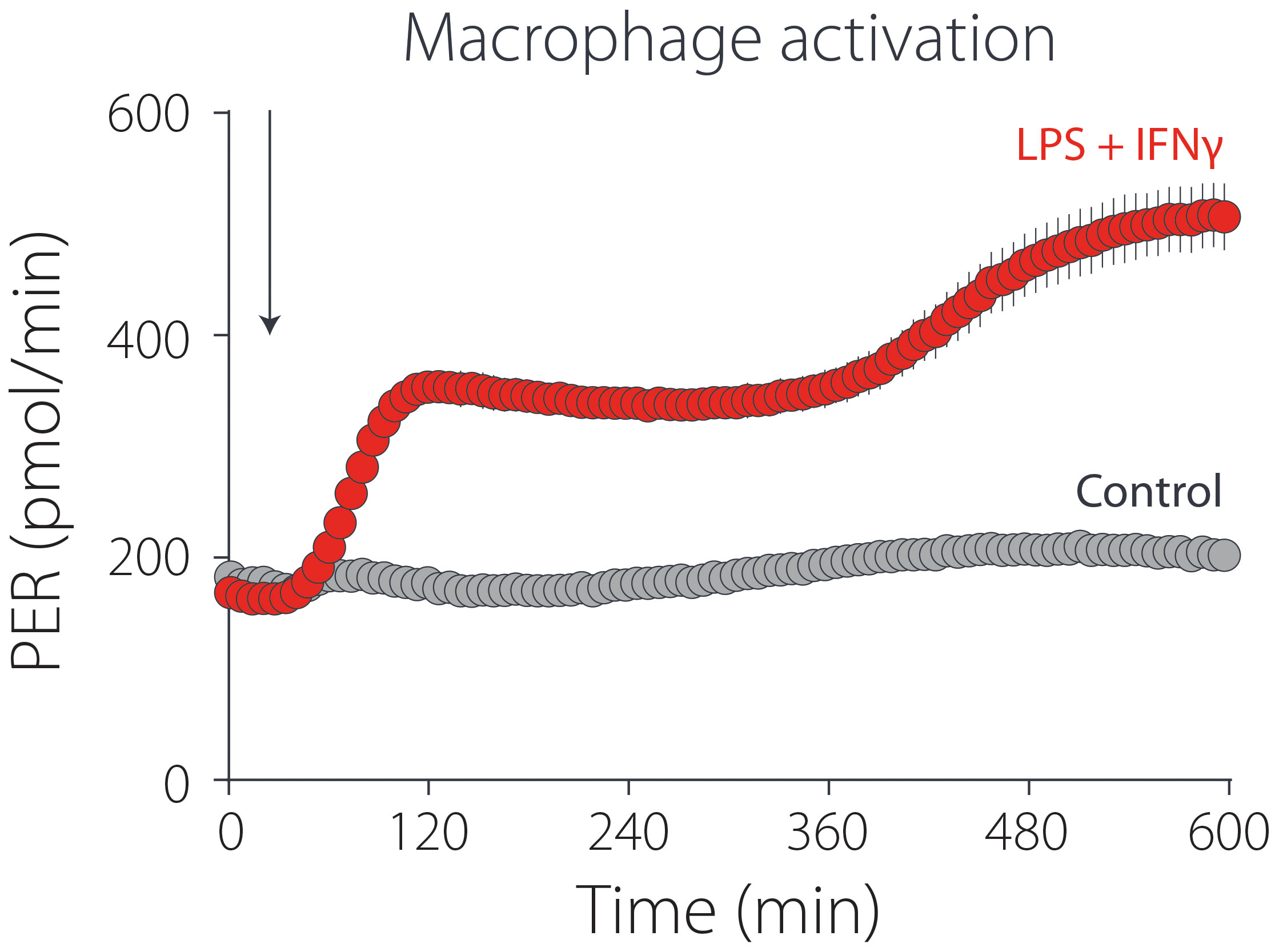
Macrophages and dendritic cells also exhibit an immediate glycolytic response to pathogenic stimuli. Similarly, as in the activated T-cell example above, glycolysis is required to support the high energy and biosynthetic demands of activated macrophages. However, in some macrophage cell types this activation is biphasic. The initial phase consists of a sustained increase in glycolytic rate with little to no impact on mitochondrial function. The second phase, occurring several hours later, is typified by a further increased glycolytic rate and a marked decrease in mitochondrial function. An example of this finding in the RAW 264.7 macrophage cell line is shown in Figure 2.
In work by the lab of Edward Pearce, Ph.D., of the Max Planck Institute of Immunobiology and Epigenetics, in Freiburg, Germany, this second phase was shown to be dependent on the production of nitric oxide.2 Nitric oxide is a well-known inhibitor of mitochondrial function, and in activated macrophages is produced by the enzyme-inducible nitric oxide synthase. Increased expression of this enzyme requires new protein synthesis—the timing of which contributes to the multiphasic nature of the response of macrophages to activation stimuli. By correlating this real-time functional information to orthogonal biological data, we have gained greater insight into the causes and metabolic implications of macrophage activation.

Figure 2. Discrimination of multiple phases of macrophage activation. Murine immortalized macrophages (RAW 264.7) were measured using an Agilent Seahorse XFe96 Analyzer. A mixture of LPS (100 ng/mL) and IFN? (20 ng/mL) were injected at the arrow. Proton Efflux Rate was monitored for an additional 9.5 hours.
Beyond Activation: Metabolic Fuels Drive Immune-Cell Fate
There are now several examples of long-lived cell types selectively choosing to oxidize fatty acids as a fuel source. While the underlying reason for this remains poorly understood, it has become clear that this “choice” to burn fat is, in fact, a requirement for the long-lived cell phenotype. Nowhere is this more evident than in the long-lived memory T-cell subpopulation that is retained following pathogen clearance. Now researchers are using this knowledge to identify mechanisms that drive T cell longevity to translate it to the clinical research setting.
CAR-T cells are T cells which have been transduced to express a synthetic cell-surface receptor. The extracellular portion of this synthetic receptor is targeted to a specific antigen of interest, and the intracellular portion is comprised of several signaling domains which translate the ligation of the external portion to intracellular activity. Recent work from the lab of Carl June, M.D., at the University of Pennsylvania has demonstrated that the intracellular signaling component is a key regulator of the long-lived central memory phenotype.3 It is not yet clear if fatty acid oxidation is supporting this long-lived phenotype in the CAR-T cells. However, there is evidence from the lab of Erika Pearce, Ph.D., at the Max Planck Institute, that fatty acid metabolism is critical not just for the function of this cell type, but for the creation of these cells in the first place.
Without Real-Time Kinetics, What Are Investigators Missing?
The vignettes highlighted here demonstrate that both short- and long-term kinetic analyses provide valuable information and mechanistic detail that would be missed by using any other approach. Researchers are increasingly focused on early events in immune-cell activation, where the response to an inflammatory signal can be tuned to impact overall cell function. In research areas such as immuno-oncology, increased activation is connected to improved cell expansion and overall cellular health; whereas in the field of immunosuppression, the converse is desired.
As the relevance of immune-cell function grows, researchers are continuously seeking tools that can enable a more refined study of the kinetics of these dynamic cell types. Agilent XF technology is uniquely poised to offer a robust, real-time view of activation, enabling the study of compounds or treatments modulating effector function.
Brian P. Dranka, Ph.D. ([email protected]), is manager of biology, cell analysis division; and Luke Dimasi ([email protected]) is manager of global product marketing and technical support, cell analysis division, at Agilent
Technologies.
References
1. P.M. Gubser, et al., “Rapid Effector Function of Memory CD8+ T Cells Requires an Immediate-Early Glycolytic Switch,” Nat. Immunol. 14, 1064–1072, doi:10.1038/ni.2687 (2013).
2. B. Everts et al., “TLR-Driven Early Glycolytic Reprogramming via the Kinases TBK1-IKK Supports the Anabolic Demands of Dendritic Cell Activation,” Nat. Immunol. 15, 323–332, doi:10.1038/ni.2833 (2014).
3. O.U. Kawalekar et al., “Distinct Signaling of Coreceptors Regulates Specific Metabolism Pathways and Impacts Memory Development in CAR T Cells,” Immunity 44, 380–390, doi:10.1016/j.immuni.2016.01.021 (2016).


