November 1, 2013 (Vol. 33, No. 19)
In the summer of 1984, Henry Erlich, Glenn Horn, Randy Saiki, and myself were working on developing a DNA test for sickle cell anemia (SCA) at Cetus Corp. SCA is an autosomal dominant disease caused by a single base mutation in the β-globin subunit of the hemoglobin gene.
Randy had developed the detection methodology for the sickle cell mutation using radiolabeled oligonucleotide probes that hybridized specifically to β-globin. These probes were cut using restriction enzymes, in a process called oligomer restriction, into two small specifically sized DNA fragments indicating the presence of normal or sickle cell alleles in the sample. These small, radioactive restriction fragments were then separated on thin-layer chromatography (TLC) plates and detected by autoradiography.
While oligomer restriction was conceptually elegant, from a practical perspective we simply could not get our probes hot enough to see a statistically significant signal above background. So, the problem we were dealing with was a classic one: how can we amplify the signal above the noise?
At that time, Kary Mullis, who conceived the idea of PCR in 1983, was running the lab that synthesized our oligo probes and was aware of the signal-to-noise problem we were having. Glenn, Randy, and I brainstormed several ideas of amplifying the signal, including using a Qβ Replicase, but none of the ideas stuck.
I remember the day in the summer of 1984 when Kary came to our lab and suggested that we try PCR to amplify the signal of the desired genes above the background of the genome.
It wasn’t until a few months later, in November 1984, that Thomas White, our vp of research, suggested to Henry that I work on reducing the PCR invention to practice as the amplification “front end” for the sickle cell anemia test.
Back then there were no thermal stable polymerases available so I started on the task of developing and characterizing the PCR methodology utilizing a fragment of E. coli DNA polymerase I (called Klenow fragment) to mediate the polymerase chain reaction. As there also were no thermal cyclers at the time, the enzyme had to be added manually at each cycle, moving the reaction tubes by hand between temperature-controlled hot plates set at the denaturation, annealing, and extension temperatures.
We had also revised the oligomer restriction assay to utilize 30% acrylamide gels instead of TLC plates to separate the now-amplified wild-type and mutant DNA fragments. The gels were dried and detected using autoradiography.
Imagine my surprise when I went to the x-ray film processor and pulled out the film of the first experiment I did in conjunction with Kary’s technician, Fred Faloona. There were two strong bands indicating the presence of the normal and mutant alleles, and virtually no noise! It had worked!
I ran to the lab and showed the results to Henry. We were very encouraged and embarked on making the PCR technology into a robust, reproducible, and repeatable methodology for use in the SCA test.
One of the most important things we did during this initial phase of PCR development was to form a team composed of Norm Arnheim, Kary, Henry, Glenn, Fred, Randy, and myself. We held meetings every Friday afternoon to review the week’s results, derive insight from the data, and decide what experiments to do next.
Success Came Quickly
From those initial laborious experiments, success built rapidly. Almost every experiment we did made the technology better and broadened the range of applications. In the summer of 1985 we wrote the first scientific paper describing the PCR methodology, and Daniel Koshland, editor of Science, had the vision to understand its significance. The first PCR paper was published in Science in December, 1985.
In the summer and fall of 1985, I cloned the human β-globin gene directly into M13 using sequence-specific primers containing restriction enzyme recognition sites. When we sequenced the ten clones, nine were β-globin, and one was δ-globin, a pseudogene. This was a major breakthrough because it meant that scientists no longer had to spend months creating bacteriophage libraries to perform molecular cloning of a gene of interest.
We published the second journal article on PCR, on its use for molecular cloning, in September, 1986 in Science.
Around this time, David Gelfand and Suzanne Stoffel at Cetus started purifying and cloning the thermal-stable DNA polymerase from the bacterial thermophile, Thermus aquaticus. This was a critical advance in the development of PCR because we did not have to add polymerase at every cycle and the thermodynamic specificity afforded by performing PCR at higher reaction temperatures resulted in a significant increase in the specificity of the amplified product.
A paper describing the use of a thermal-stable DNA polymerase was published in Science in 1988, and PCR as a major breakthrough in the history of molecular biology was really off and running.
Real-time PCR and Taqman™ technology were also originally conceived by the PCR team at Cetus. Russell Higuchi had the insightful realization that by using fluorescence signals to track the exponential growth curve of PCR amplicons during the reaction, this information could be used to quantitate the amount of DNA in a sample. Real-time PCR was born.
Randy, a fan of the Pac Man™ video game, conceptually piggy-backed off real-time PCR with the Taq Man methodology of using a PCR product-specific, fluorescently labeled oligo probe that would be detected only when cleaved during the amplification process.
The Taqman chemistry, as it is known today, was eventually reduced to practice by Linda Lee at Applied Biosystems (now Life Technologies). PCR has been instrumental in the development of DNA sequencing, initially with cycle sequencing, which greatly facilitated Sanger sequencing to the present day, where PCR is the key step in generating the libraries required for next-gen sequencing.
PCR continues to evolve and improve. Digital PCR enables one to quantify the amount of DNA or nucleic acid in samples with hitherto unachievable level of accuracy and precision. It could become a critical methodology in cancer diagnostics for monitoring minimal residual disease.
With respect to infectious disease and genetic testing, PCR has become the engine that drives the molecular diagnostic industry. Because of the relatively low cost for the amount of information that can be obtained per test, it has enabled diagnostic testing ranging from HIV to SARs to screening newborns for cystic fibrosis.
Stephen Scharf is a senior staff scientist with Life Technologies, the developer of the QuantStudio™ 3D Digital PCR System and TaqMan® Mutation Detection Assays.

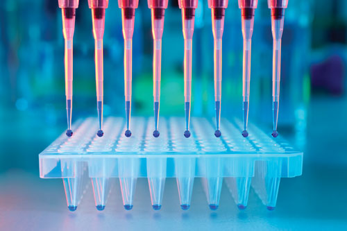
PCR did for molecular biology what the transistor did for electronics. It enabled an ever-broadening range of applications, starting with molecular cloning. [anyaivanova/iStock]
Stepping Past Taq
The advent of PCR revived interest in thermophilic DNA polymerases, building on earlier studies of Taq DNA Polymerase. Coincident discoveries of extremely thermophilic microorganisms in ocean thermal vents, growing at temperatures near 100°C, provided an alternate source for even more thermostable enzymes. One such organism, Thermococcus litoralis, yielded an extremely thermostable DNA polymerase, Vent® DNA Polymerase, the first of many DNA polymerases displaying enhanced thermostability relative to Taq DNA Polymerase, and additionally possessing enhanced fidelity due to an inherent proofreading activity.
Cloning the Vent DNA Polymerase gene presented a challenge: DNA sequencing revealed an encoded peptide of 180 kDa, twice the size predicted from protein gels. This discrepancy was resolved by identification of two auto-catalytic post-translational protein splicing events, a novel concept at the time. Reconstructing the gene without intervening protein sequences was critical to high-yield polymerase expression.
This DNA polymerase is a member of a highly homologous family of archaeal DNA polymerases. Family members have been extensively engineered and optimized by many groups, and have played essential roles in the expansion of PCR protocols at the heart of workflows including cloning, site-directed mutagenesis, and library preparation, as well as SNP detection and an expanding array of isothermal amplification applications.
Bill Jack, Ph.D. ([email protected]) is the research director at New England Biolabs.


