April 1, 2009 (Vol. 29, No. 7)
ELISA-Based Method Simplifies Process and Provides Results in HTS Format
Epigenetics is often defined as the study of heritable changes that occur without a change in the actual DNA sequences. It has been shown that associated DNA proteins, called histones, are altered by methylation, phosphorylation, acetylation, and other changes. DNA chromatin structure is altered by these histone modifications as well as by direct methylation. These changes are in constant flux and can alter gene expression and related phenotypes; the actual DNA sequence, however, remains unchanged.
DNA methylation, the addition of a methyl group to the 5´ carbon of cytosine, is one of the main components of the epigenetic code and is thought to be involved in the repression of gene activity. It’s relationship to development and cancer has been extensively studied. While it is clear that epigenetic regulation is complex, a simplified model starts with double-stranded DNA wound around a set of completely acetylated histones and starting as an activated, fully transcribed gene.
Transcriptional repression can be initiated near the promoter by deacetylating specific lysine residues of nearby histone proteins. Subsequently, the lysines can be methylated up to three times per lysine, each time locking in gene shutdown. Finally, the cytosines within the gene can be methylated at its 5´ carbon to further repress the gene. Although the progression is complex, with some histone methylation actually activating DNA transcription, one may think of this general progression of changes as a compaction of the gene into dense, untranscribable chromatin.
Recent emphasis has been given to CpG island methylation of the promoter region of specific genes. There are others interested in the overall or global methylation of the entire genome, however. Global methylation varies from diseased to normal states, from tissue to tissue, between genders, and as we age. It is hoped that global methylation profiling can lead to disease-state biomarkers and early diagnosis.
Several methods exist to measure global methylation levels. Immunohistostaining is commonly used to look at tissue samples, but it has low sensitivity. There are several techniques that use methylation-specific restriction enzymes, but the protocols can be lengthy and cumbersome and often include the use of radioactivity.
LC-mass spectrometry is considered the gold standard for DNA-methylation analysis as it provides the user with the exact amount of methylated cytosines in a sample. It is costly, however, and requires a certain amount of expertise, and the DNA must be digested to the single nucleotide level prior to analysis. Recently, Sigma Aldrich created a global methylation analysis method, similar to a sandwich ELISA that enables the direct quantitation of genomic DNA methylation. The kit takes four hours, and is amenable to a high-throughput format and friendly to biological researchers.
Workflow
Purified genomic DNA is hybridized to assay wells, which are then blocked and subsequently probed by a capture antibody specific for the methylated cytosines in DNA. After a wash step, detection is facilitated using an enzyme linked antibody, and the amount of this enzyme is quantified colorimetrically after washing away excess. The samples are read at A450 on a standard plate reader; the entire process takes approximately four hours.
Experimental Results
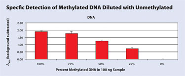
The ability to distinguish between methylated and unmethylated DNA was the first obstacle this technique had to overcome.To create a known methylated sample, unmethylated Lambda DNA was treated with SssI Methylase and then purified. Both treated and untreated DNA, in the amount of 100 ng, were added to assay wells in triplicate and analyzed as indicated above. The untreated sample was comparable to the blank indicating no methylation present. The treated sample had a sixfold increase in signal indicating that methylation occurred due to treatment with the methylase (Figure 1).
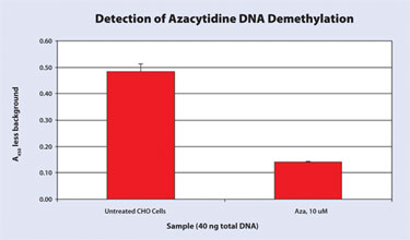
The next experiments were designed to prove that the kit would work with DNA purified from a biological system. These tests took advantage of azacytidine, which is a chemical analog of cytidine that cannot be methylated. When added to cell culture media it is incorporated into DNA during replication and thus creates DNA with a decreased global methylation. Azacytidine was added directly to the media of CHO DG44 cells. The cells were harvested after 48 hours. Global methylation was measured on 40 ng of the purified CHO DG44 DNA in triplicate from treated and untreated cells. The treated cells had a threefold decrease in methylation due to the azacytidine treatment (Figure 2).
DNA hypomethylation is of particular interest to cancer researchers. In cancer, hypomethylation is seen in heterochromatic DNA repeats, dispersed retrotransposons, endogenous retroviral elements, and unique sequences such as transcription control sequences. It is thought that these events occur early in the tumorigenesis process, and global hypomethylation could be a potential biomarker.
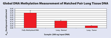
DNA from matched tissue and tumor samples was evaluated with our method to determine if shifts in global methylation could be detected. DNA was isolated from bronchioalveolar carcinoma and adjacent normal lung tissue from a 72-year-old male. Global DNA methylation was measured for both samples and compared to a fully methylated human genomic DNA control.
As expected, the adjacent normal lung tissue was less methylated than the fully methylated control and the tumor DNA was hypomethylated when compared to normal lung tissue (Figure 3). In a second experiment, DNA was isolated from hepatocellular carcinoma and adjacent normal liver tissue from a 54-year-old male.
Global DNA methylation was measured for both samples and compared to a fully methylated human genomic DNA control. The normal liver DNA was slightly less methylated than the control. Previous publications indicate that hepatocelluar carcinoma is hypomethylated. Our data confirms that tumor liver DNA is hypomethylated in this comparison.

Figure 1. Various mixtures (100 ng) of methylated and unmethylated DNA were prepared and global methylation was measured.

Figure 2. Azacytidine, a known demethylating agent, was added directly to the media of CHO DG44 cells.

Figure 3. DNA was isolated from bronchioalveolar carcinoma and adjacent normal lung tissue from a 72-year-old male.
Future Work
While the Imprint Methylation DNA Quantification kit is useful in distinguishing relative levels of global DNA methylation, LC-MS is considered the gold standard for making absolute measurements. Work is under way to convert this relative, ELISA-based method to an absolute quantitation technique that will be comparable to LC-MS. Samples to be analyzed include SssI methylated DNA, normal human genomic DNA, tumor DNA, and DNA from various cell lines. The data generated from these comparisons is expected to provide a correlation coefficient between LC-MS analysis and our method.
Conclusion
Until now, measurement of global DNA methylation could be cumbersome and often, cost prohibitive. We have demonstrated that our ELISA-based method, the Imprint Methylated DNA Quantification kit, simplifies the process and provides results in about half a day in a high-throughput format. Our method easily distinguishes between methylated and unmethylated DNA.
Methylated DNA can be detected in a mixed population of methylated and unmethylated DNA. The signal generated by this method is proportional to the amount of methylated DNA in the sample. DNA from biological systems such as cultured cells, normal tissue, and tumors have also been analyzed. The global methylation levels determined with our method correspond with data generated by other methodologies.
Deborah L.Vassar is R&D scientist, Ernest J. Mueller, Ph.D., is R&D principal investigator, and Savita Bagga, Ph.D. ([email protected]), is product manager, epigenetics at Sigma-Aldrich. Web: www.sigmaaldrich.com/life-science/molecular-biology/epigenetics.html.


