In the late 1960s, Bonucci and Anderson first reported finding small, secreted extracellular vesicles in bone cells. Over the next 20 years, through the combined use of electron microscopy, ultracentrifugation, and functional studies, other types of cells including cancer cells and immature red blood cells were also found to secrete extracellular vesicles with great biological significance, and this field has grown substantially.
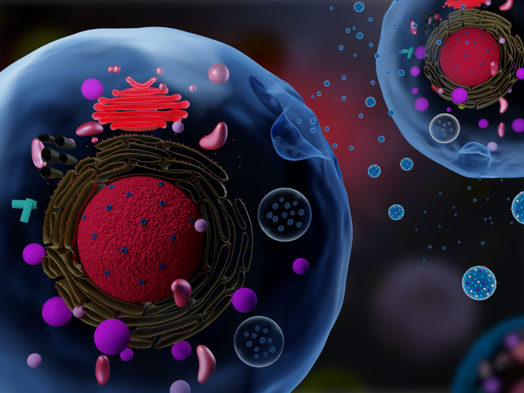
There are two main types of extracellular vesicles. The bigger submicron-sized ones are called microvesicles while those that are between 30–150 nm in diameter are collectively referred to as exosomes. More specifically, exosomes are a type of nanometer-sized extracellular vesicles that originate through budding from the plasma membrane and endosome membrane. They contain a variety of cargo, including nucleic acids, lipids, and proteins and express transmembrane proteins and receptors that facilitate intracellular communications.
Over the last six to seven decades, research has shown that exosomes play crucial roles in human health and biology, including regeneration and cancer metastasis. Additionally, there have been innovative attempts to exploit exosomes for diagnostics and as agents for biomolecule delivery. Here, we will focus on existing challenges and emerging technology to engineer and translate exosomes for clinical use.
Low throughput, yield, and purity in exosome isolation
Ultracentrifugation is currently the most popular method for exosome isolation but this method suffers from low throughput and purity, especially from contaminants like cell-free nucleic acids and lipo-proteins present in samples. A variety of other methods using mechanisms based on precipitation, affinity, and size have also been developed although with limited success.
“Extracellular vehicles (EVs) and exosomes have shown great potential in disease diagnostics and therapeutics. One of the major challenges for clinical translations is EV isolation. We introduced negative pressure oscillation and double-coupled harmonic oscillator–enabled membrane vibration for removing free proteins and nucleic acids even from the high-concentration EVs. We named this technique exosome detection via the ultrafast-isolation system (EXODUS) for speedy and clog-free purification of exosomes,” says Fei Liu, PhD, professor at the Wenzhou Institute, University of Chinese Academy of Sciences.

EXODUS applies double coupled harmonic oscillations into a dual-nanoporous membrane filter system to generate transverse waves to remove small particles like nucleic acids and proteins while trapping bigger-sized exosomes. The oscillations also reduce the probability of membrane fouling which is known to inhibit permeated flux, thus enhancing exosome isolation speed, yield, and purity. This method can be used to prepare EVs for downstream analysis such as polymerase chain reaction and next-generation sequencing.
The researchers compared EXODUS with other commonly used exosome isolation techniques, including ultracentrifugation, poly-ethylene-glycol precipitation, and size-exclusion chromatography and showed that EXODUS outperformed these methods in terms of relative purity, relative yield, and processing time. Most notably, by optimizing the frequency of the harmonic oscillations, exosome isolation which can take up to 12 hours can be completed in as fast as 0.16 hr with EXODUS.
Using western blot analysis, Yuchao Chen, a PhD student at the Wenzhou Institute, University of Chinese Academy of Sciences, and team also demonstrated that exosomes isolated from urine samples with EXODUS had significantly higher concentration of major exosomal urine proteins, including Alix, CD63, TSG101, and CD81 while being low in uromodulin (UMOD), a major protein contaminant in urine. EXODUS also isolated exosomes with numbers linearly correlating with different urine volumes, showing that this technique is stable and useful over a wide dynamic of workable sample volume and concentration range.
Finally, using EXODUS, the team collected urine samples from patients with bladder cancer, kidney cancer, and patients with noncancerous urinary diseases as control participants. Exosomal transcriptome revealed that in bladder cancer, but not kidney cancer, genes encoding for energy and amino acid metabolism and protein synthesis and processing regulation were selectively upregulated. This finding demonstrates the utility of EXODUS as a sensitive, ultrafast exosome purification method that may reveal metabolic reprogramming pathways associated with cancer development for diagnostics or monitoring of therapeutic interventions.

To capitalize on the use of exosomes for clinical applications, it is also useful to integrate exosome isolation method with downstream detection and analysis techniques. Sijun Pan, PhD, of the Institute for Health Innovation & Technology, National University of Singapore, and colleagues recently developed a novel method coined extracellular vesicle monitoring of small-molecule chemical occupancy and protein expression (ExoSCOPE) for measuring activity and efficacy of drug treatments against tumors by characterizing extracellular vesicles in patient blood samples. This technique, which makes use of probe amplification and spatial patterning of molecular reactions within plasmonic resonators, is a powerful example of how the use of extracellular vesicles such as exosomes can be exploited for clinical applications.
Poor control of intra-exosomal content
While exosomes are attractive candidates for drug delivery as they exhibit properties such as tissue tropism and can be engineered to bind cells expressing specific receptors, they contain a variety of biomolecules such as nucleic acid and proteins inside of them that may not be therapeutically useful. Genetic approaches have been reported to create cells to enhance production of designer exosomes but this method requires complex genetic modifications that add a layer of complexity for clinical translation.
Recently, Lemmerman, et al. made use of a nonviral nano-transfection method that provided electrical forces through nano-scale structures to electroporate fibroblasts and electrophoretically inject plasmids encoding for developmental transcription factor genes Etv2, Foxc2, and Fli1 (EFF) into primary mouse embryonic fibroblasts.
Using quantitative reverse transcription polymerase chain reaction (qRT-PCR) analyses, the team found that the nano-transfected fibroblasts significantly increased production of EFF proteins compared to un-transfected controls. Following which, they showed that the fibroblasts transfected with EFF plasmids produced exosomes loaded with provasculogenic and angiogenic EFF transcripts to drive reprogamming-based vasculogenesis as a potential therapy for ischemic stroke.
In addition, mice injected with EFF-nano-transfected fibroblasts exhibited improvement in ischemic stroke-related locomotive impairment. Immunohistological examinations also showed that treated mice had higher brain vascularity and reduced glial scar formation. The nonviral nano-transfection method is a promising strategy to modulate content of exosomes for use in clinical therapies.
Poor scalability in exosome production
Despite the advantages of exosomes for biomedical applications, it is important to recognize that to translate them for clinical applications, they need to be produced in sufficient quantities. Currently, this is still a challenge because not many cells secrete sufficient numbers of exosomes.
Making use of a similar mechanism to the aforementioned nano-transfection termed cellular nanoporation (CNP), Zhaogang Yang, PhD, a former Ohio State postdoctoral researcher now at the University of Texas Southwestern Medical Center, and co-workers electrically enhanced the production of exosomes with 50-fold larger than bulk electroporation and methods like hypoxia, starvation, and heat treatment that induce global cellular stress responses. The magnitude of voltage applied with CNP can also be optimized for exosome production.
To understand how nano-electrical stimulation enhanced exosome production, the authors made use of transmission electron microscopy and found that CNP significantly increased the formation of multivesicular bodies by two-fold and interluminal vesicles by eight-fold. Nano-electrical stimulation also led to calcium ion influx into cells to trigger exosome release. Finally, electrical stimuli might have also induced heat-shock response that promoted the p53–TSAP6 signaling, leading to increased exosome production as a part of the cellular recovery process.
“mRNA-based therapy has a great potential for treating many undruggable diseases for unmet medical needs in areas such as regenerative medicine and cancer treatment. Even though synthetic mRNA is available in recent years, its delivery using synthetic nanoparticles has many drawbacks including poor efficacy to pass physiological barriers such as blood-brain barrier, toxicity of some synthetic components, and low targeting and cellular uptake in vivo. Viral vectors can deliver mRNA efficiently in vivo, but unpredictable and severe immune responses are major concerns,” says James Lee, PhD, professor of chemical & biomolecular engineering at the Ohio State University.

“Extracellular vesicles including exosomes, on the other hand, are safer than viral vectors and more efficient than synthetic nanoparticles. However, loading large and unstable biomolecules such as mRNAs in tiny exosomes is very difficult. We tried to solve this problem by using our cellular nanoporation platform to enhance exosome production and load endogenous mRNAs (and noncoding RNAs) in secreted exosomes simultaneously.”
Other physical fields including mechanical forces have also been found to increase production of extracellular vesicles like exosomes. “This 2D culture condition is less physiologically relevant, and the production usually involves large volumes of medium, space, and intensive labor. The goal of our study was to overcome the low EV yield from current practices. Current EV isolation remains largely from parental cells cultured as monolayers on 2D platforms. We adopted a tissue engineering approach, by applying two forms of mechanical stimulations (i.e., via flow or stretching) on multiple engineered tissues inside bioreactors, aiming to boost EV secretions to meet clinical demands,” says Shulamit Levenberg, PhD, professor of biomedical engineering at the TECHION–Israel Institute of Technology.

Guo, et al. grew 3D tissues containing stem cells or muscle cells in bioreactors for flow stimulation or mechanical stretching and stimulated tissues produced up to 37-fold more extracellular vesicles than static controls. To understand the mechanism behind mechanical force-induced exosome production, the team characterized the expression of yes-associated protein (YAP) that is linked to nuclear transduction of mechanical cues. They found that stimulated cells expressed up to 6-fold higher YAP proteins than static controls. In the presence of verteporfin, a YAP inhibitor, production of extracellular vesicles was significantly quenched.
“We were able to significantly scale up extracellular vesicles production (~150-fold increase) with our method and showed that it was mediated by YAP. One limitation is that we did not examine the therapeutic effects of mechanically stimulated EVs in relevant disease models, but our study demonstrated the possibility to harness commercial bioreactor systems in pharmaceutical companies to generate sufficient extracellular vesicles for clinical use,” noted the researchers.
Outlook
Exosomes have shown great potential as a type of biocompatible biological material for biomolecule delivery and diagnostics for a variety of applications like tissue regeneration, drug screening, and cancer treatment. However, to be useful clinically, better technologies are needed to isolate exosomes in a fast manner with high yield and purity. Additionally, techniques to control intra-exosomal content and boost exosome production from cells would benefit clinical translation.


