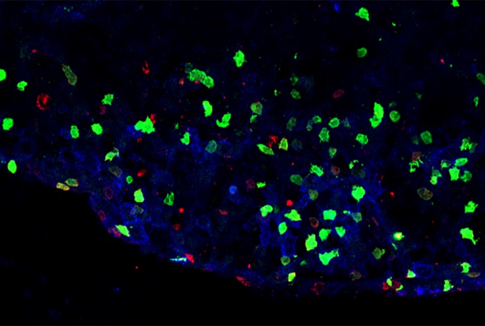
A new mouse study by researchers at Weill Cornell Medicine and New York-Presbyterian demonstrates a group of immune cells that protect against inflammation in the gastrointestinal tract may have the opposite effect in multiple sclerosis (MS) and other brain inflammation-related conditions.
Their findings are published in the journal Nature in a paper titled, “Antigen-presenting innate lymphoid cells orchestrate neuroinflammation.”
“Pro-inflammatory T cells in the central nervous system (CNS) are causally associated with multiple demyelinating and neurodegenerative diseases, but the pathways that control these responses remain unclear. Here we define a population of inflammatory group 3 innate lymphoid cells (ILC3s) that infiltrate the CNS in a mouse model of multiple sclerosis.”
The researchers set their sights on studying a set of immune cells called ILC3s, which are key sentinels of barrier tissue homeostasis and respond rapidly to damage, inflammation, and infection to restore tissue health.
The researchers located a unique subset of these ILC3s that circulate in the bloodstream and can infiltrate the brain, and surprisingly promote inflammation. The scientists called this subset inflammatory ILC3s, and found them in the central nervous system of mice with a condition modeling MS.
“This work has the potential to inform our understanding of, and potential treatments for, a broad variety of conditions involving T-cell infiltration of the brain,” explained senior author Gregory Sonnenberg, PhD, associate professor of microbiology and immunology in medicine in the division of gastroenterology and hepatology and a member of the Jill Roberts Institute for Research in Inflammatory Bowel Disease at Weill Cornell Medicine.
“The infiltration of these inflammatory ILC3s to the brains and spinal cords of mice coincides with the onset and peak of disease,” said first author John Benji Grigg, a Weill Cornell Graduate School of Medical Sciences doctoral candidate in the Sonnenberg laboratory. “Further, our experimental data in mice demonstrate these immune cells play a key role in driving the pathogenesis of neuro-inflammation.”
The researchers found that they could prevent MS-like disease in the animals by removing from the ILC3s a key molecule called MHCII. The removal of MHCII blocks the cells’ ability to activate myelin-attacking T cells.
“Despite our very best disease-modifying therapies for MS, patients continue to progress, and since disease onset is early in life, they face the prospect of permanent physical and cognitive disability,” said co-author Tim Vartanian, PhD, professor of neuroscience in the Feil Family Brain and Mind Institute at Weill Cornell Medicine, chief of the division of multiple sclerosis and neuro-immunology and a professor of neurology in the department of neurology at Weill Cornell Medicine and NewYork-Presbyterian/Weill Cornell Medical Center. “Identification of inflammatory ILC3s with antigen presentation capabilities in the central nervous system of people with MS offers a new strategic target to prevent nervous system injury.”
Finally, the researchers discovered that ILC3s that reside in other tissues in the body can be programmed, in effect, to counter the activity of brain-infiltrating T cells, preventing the MS-like condition disease in mice.
“Collectively, our data define a population of inflammatory ILC3s that is essential for directly promoting T-cell-dependent neuroinflammation in the CNS and reveal the potential of harnessing peripheral tissue-resident ILC3s for the prevention of autoimmune disease,” concluded the researchers.


