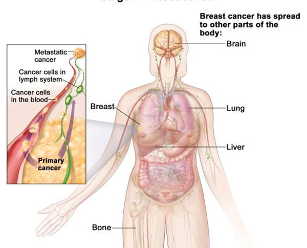
The spread of breast cancer to tissues and organs distant from the original tumor site is associated with poorer patient prognosis, and anticancer therapies are generally far less effective against metastatic disease. However, the mechanisms controlling metastasis aren’t well understood. Scientists in the U.S. and Australia have now discovered that the primary tumor can influence whether metastasis-initiating cells (MICs) that break away from the primary tumor site will be able to colonize new environments in the body.
Studies in mice led by a team at Brigham and Women’s Hospital, Harvard Medical School, and the Garvan Institute of Medical Research, showed that some primary breast tumors can trigger inflammatory reactions at distant sites, which essentially freeze the MICs in an intermediate state and prevent them from differentiating into cells that can form new tumors. The researchers also found that higher levels of the same inflammatory signals were associated with better overall survival and metastasis-free survival in metastatic breast cancer patients, suggesting that the interaction between the primary tumor and MICs could represent a target for anticancer therapy.
“Our findings flip the current thinking on its head,” suggests Sandra McAllister, Ph.D., a researcher at the division of hematology at Brigham and Women’s Hospital. “Many people study primary tumors and the assumption has been that metastases grow the same way. But our work suggests that, while inflammation can help tumor cells escape and land elsewhere in the body, if inflammation is there when they land, it keeps the cells in check. When inflammation is suppressed, the cells grow out.” Dr. McAlister is co-senior author of the researchers published paper in Nature Cell Biology.
“This new research has yielded that rare thing — a clue from the cancer itself about new possibilities to fight its spread,” adds co-senior author Christine Chaffer, Ph.D., at the Garvan Institute. “Our goal is to work out how we can mimic this 'freezing' of secondary cancers, so that one day we might influence all breast cancers to keep their secondary tumors in check.” The team reported its findings in a paper titled, “IL-1β inflammatory response driven by primary breast cancer prevents metastasis-initiating cell colonization.
Although most breast cancer patient are diagnosed before they show evidence of metastasis, up to 30% of patients will develop metastatic disease, even if not until decades later. In fact, the process by which cancer cells break away from the primary tumor site and travel to distant tissues and organs often begins early, the authors note, but not of these escaped cells will actually seed new tumors. “By some estimates, less than 0.02% of those disseminated tumor cells will form secondary tumors, indicating that successful metastatic colonization is rare and ascribed to only specialized minority cancer cells, termed MICs. What scientists don't yet understand is the process by which MICs can exploit their cellular plasticity to generate metastatic lesions.”
The researchers’ studies in a mouse model of metastatic breast cancer have now demonstrated that primary tumor cells can trigger a systemic inflammatory response involving IL-1β-secreting innate immune cells, which reaches distant MIC microenvironments and freezes the MICs in a transitional state, preventing them from developing into new tumors in the lung and other tissues. When the researchers analyzed secondary tumors in the lungs of mice carrying either metastasis-inhibitory primary human tumors or noninhibitory primary tumors, they found that suppression of the MICs was associated with high levels of neutrophils and macrophages, and upregulation of IL-1β. Knocking down neutrophils from the secondary tumor environments was associated with the development of significantly larger pulmonary metastases. In contrast, delivering IL-1β to tumors was enough to trigger macrophage infiltration, and the development of a tumor-promoting MIC phenotype, “demonstrating that a single dose was sufficient to trigger a sustained inflammatory response and maintain a mesenchymal phenotype.”
The results indicated that innate immune cells secreting IL-1β, and mobilized by the primary tumor, could block MIC colonization in the mouse model by preventing them from differentiating into active tumor cells, but only if the inflammatory signals were present at the secondary site before the MICs started to differentiate. Secondary tumors were not inhibited if the IL-1β-dependent inflammatory cascade was instigated after MIC dissemination and growth initiation at the distant site.
“When breakaway cells are forced to remain in the transition state, they don't grow very well,” remarks Dr. McAllister “and their ability to form a new tumor is severely compromised. So, remarkably, by activating the immune response, the primary tumor essentially shuts down its own spread.”
To see whether an equivalent relationship between inflammatory response and metastasis development occurs in humans, the team analyzed gene expression data to check for IL-1β expression among breast cancer patients who had either lymph node-positive, or lymph node-negative disease. The results found no relationship between IL-1β expression and overall survival among 508 patients with lymph node-negative disease. In contrast, among 215 patients with lymph node-positive disease, high IL-1β expression was associated with higher overall survival compared with patients with low IL-1β.
The researchers suggest that their results indicate both that MICs are specifically susceptible to growth inhibition, and that “therapies designed to prevent disseminated MIC differentiation compromise their ability to form lethal metastases.”
The results also indicate that proinflammatory processes that may be involved in cancer initiation may conversely hold back the development of metastases. “Inflammatory processes that initiate primary disease and drive EMT [epithelial-to-mesenchymal transition] in primary breast cancers, thus causing MICs to disseminate, are not necessarily productive for MIC colonization,” they write. “… by specifically examining the role of IL-1β after MIC dissemination, we learned that sustained IL-1β-mediated inflammation or MIC IL-1R signaling prevents colonization and must be shut down for secondary tumor formation. We, therefore, consider that IL-1β has both dissemination-supportive and colonization-suppressive functions.”
One of the implications of the findings is that removing the primary tumor could actually support the development of metastasis by taking the brakes of secondary tumor development by circulating MICs. “It is, therefore, reasonable to think that a primary tumor expressing pro-inflammatory cytokines resulting in activation of an IL-1β-dependent innate immune response in the metastatic niche might keep secondary disease at bay and conversely, that primary tumor removal might prompt recurrence,” they concluded. “Our data reveal complex interactions that occur between primary tumors and disseminated MICs that could be exploited to improve patient survival.”
“An important implication of our study is that therapies that may prevent the growth of primary tumors may not confer the same beneficial effects for preventing the growth of metastases and further research is required,” Dr. McAllister concludes.
The researchers hope that their findings could ultimately help to new approaches to preventing metastasis in breast cancer, but also potentially in other tumor types. “We want to understand exactly what the tumor is releasing to activate this immune response, and how immune cells are targeting the secondary sites,” adds Dr. Chaffer. “In principle, all of these steps present therapeutic opportunities that could be used to stop a cancer from developing any further.”


