
January 15, 2016 (Vol. 36, No. 2)
Cell-based assay design has been overshadowed recently by an uptick in other omics solutions. It still plays an important role, however, in the diagnosis of disease, in the analysis of gene products, and in the study of drug efficacy and toxicity.
To better predict the changes that occur in physiological systems, companies are investing in the development of 3D systems. Companies hope that improvements in 3D systems will more accurately inform our understanding of cell-cell interactions and pharmaceutical efficacy.
Not everyone is convinced that game-changing improvements are imminent. Regardless, many contract research organizations (CROs), life sciences companies, and clinical research laboratories are beginning to embrace 3D system improvements such as microscaffolds and 3D matrices. Indeed, these methods are already playing an important role in cell-based assay development.
One CRO focused on combining advances in culture methods with preexisting cell-based assay technology is Pelago Bioscience. The company’s CEO, Michael Dabrowski, Ph.D., believes that future cell-based assays will be cell culture systems that display intracellular signaling cascades mimicking the in vivo situation. “Phenotypic screening with patient-derived cells or induced pluripotent stem cells,” explains Dr. Dabrowski, “are playing an increasingly prominent role in the discovery of novel therapeutic candidates.”
Other mimics of in vivo systems are also on the rise. “High-content screening on 2D and 3D organotypic cultures or tumor spheroids,” notes Dr. Dabrowski, “will be important and enable testing of novel therapeutic approaches.” Nonetheless, Dr. Dabrowski believes that we still need to fully understand the added value of these more complex systems, their reproducibility and relevance, before we discard in vivo testing.
Cellular Thermal Shift Assay
Pelago Bioscience, a spinout from the Karolinska Institute in Stockholm, was founded around the patented Cellular Thermal Shift Assay (CETSA), which allows for direct measurements of molecule and target protein interactions by exploiting a known shift in melting point caused by small molecule interactions. CETSA can be readily combined with many other technologies—including cultures of cells, tissues, and organs—to determine target engagement “without modifications to the target or compounds investigated,” says Dr. Dabrowski.
GlaxoSmithKline (GSK), a CETSA license holder, has been using CETSA along with mass spectrometry to profile its candidate drugs on 4,000–8,000 cellular protein targets for on- and off-target effects. This technology has been very useful in the analysis of both preclinical and clinical samples such as patient-derived tumors—not that CETSA’s sample analysis capabilities are limited to cancer studies. “The CETSA method,” asserts Dr. Dabrowski, “is readily applied on more than half of the druggable proteome,” including proteases, enzymes, and kinases, suggesting that CETSA could benefit customers “in all therapy areas.”
Microscaffolding for Cell-Based Assays
“Clients tend to be very conservative until you show them data specific for their project,” says Gary Allenby, Ph.D., CSO and cofounder of Aurelia Bioscience. He has worked with a variety of developing culture systems and currently has a grant to design a 3D cell culture system using microscaffolds and electrospun materials. In his microscaffolds, adherent cells change the way they grow and divide.
“Over 80% of the assays we run at Aurelia Bioscience are cell based,” Dr. Allenby points out. “Although many remain recombinant, we strive to implement the testing of compounds in more physiological environments.”
“We are firm believers,” he says of his team at Aurelia Bioscience, “in phenotypic drug discovery.” Dr. Allenby explains that his team focuses on the biological mechanisms of systems. Further, Dr. Allenby suggests that focusing on the “interplay between cells as part of a mixed population” enables his team to develop and improve biologically relevant approaches for microwell-based cellular assays.
“The only problem,” he complains, “is that everyone is putting cells from 2D systems—cells that have been cultured over tens of years—into 3D systems. How can the same cell, when cultured under the equivalent of 1,000 feet of media, possibly respond normally? I think I would look and behave differently [if I were subjected to a comparable change]. I just don’t see how studying things out of context can help the situation.”
The potential for context explains why Dr. Allenby and others in the field are excited about 3D systems. These systems are, among other things, aimed at improving blood flow and oxygen levels and creating more cost-effective scaling for cell-based technologies.
“Each format has advantages and disadvantages,” he argues. “The next 10 years will see a plethora of technologies that use bio-inks, scaffolds, and new reagents” to develop improved models of biological systems. These models will be custom tailored for compound screening, specific cells, and specific readouts.
“Our collaborators,” confides Dr. Allenby, “want to use these models for regenerative medicine purposes.” Yet Dr. Allenby insists that he is interested in screening compounds for clients using his label-free approach to ligand-receptor measurement.
Development of 3D Culture Technology
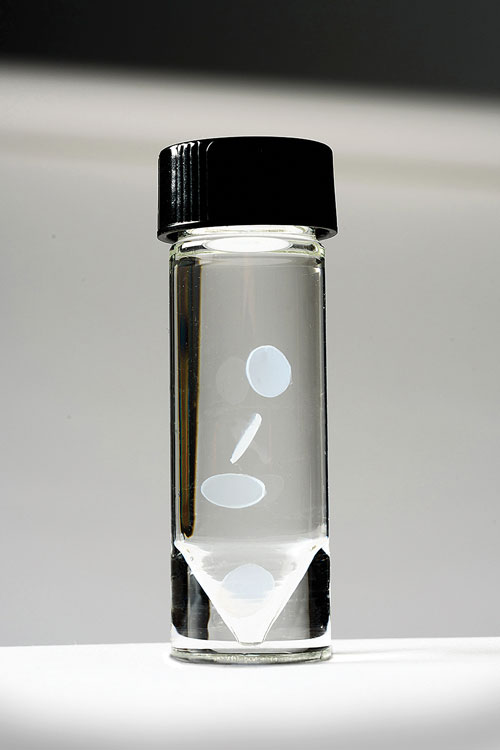
One system currently on the market is the RAFT™ 3D Culture System. It was developed by TAP Biosystems, and it has been distributed by Lonza Bioscience since July 2015.
The RAFT System combines rat tail collagen type 1 with a patented absorber to create a more realistic cellular growth system. In its first incarnation—as per the original University College London design—the RAFT System included a compression system “to condense the collagen to a high density structure,” says Lubna Hussain, senior product manager for 3D culture and primary cells at Lonza Bioscience. TAP Biosystems then bought the technology.
“The company further developed that concept into an actual product,” she continues, “with simple absorbers to remove the liquid rather than using a more complex, multicomponent pressure system.” The company also expanded the system through kit development, which provided “everything users needed”—except cells, specific lab media, and pipet tips.
Hussain and others at Lonza Bioscience have looked at market reports that suggest significant growth in the 3D culture market over the next five years. Better modeling of in vivo systems in a 3D culture format and the desire to reduce animal testing are playing an important role in this growth trajectory.
According to Hussain, 2D systems are not the ideal solution. Instead, people are turning to 3D systems in hopes of improving the biological relevance of cell culture assays. These positive growth trends and Lonza Bioscience’s expertise in this market segment fueled the company’s decision to distribute the RAFT system.
“We already sell primary cells, media, and transfection devices,” informs Hussain. “So, essentially, this was another way for our customers to elevate their research.”
Application of 3D Culture Technology
The RAFT System and other 3D culture systems can be used for a variety of cell-based assays including immunocytochemistry, viability, and toxicity analysis. “The RAFT cultures are condensed to a thickness of about 100–120 μm,” says Jenny Schroeder, Ph.D., a senior scientist at Lonza Cologne. Such cultures, she says, are highly transparent, allowing for fluorescent-based dye visualization.
The RAFT System is a hydrogel, similar to other extracellular matrix products on the market, such as Matrigel, sold by Corning. Dr. Schroeder points out, however, that Matrigel consists of laminin, collagen type 4, and entactin, “reflecting the basement membrane, a thin ECM layer in vivo,” whereas the RAFT System forms a 3D structure that is similar to that found in human tissues. The RAFT System’s structure, she adds, is compatible with a broad variety of assays including fluorescence microscopy.
One part of Lonza’s internal work with the RAFT System is to examine differences in cell growth in a RAFT model of the lung. One of the reasons for interest in more advanced culture systems, such as the RAFT System, Dr. Schroeder says, is that these systems can be used with human cells to get data of higher biological relevance. “For example,” notes Dr. Schroeder, “in a lung model, cells derived from healthy and asthmatic donors can be compared to see if there is a difference in how the cells behave.” Results to this effect were cited by Lonza during a presentation at the 2015 American Society for Cell Biology conference.
Establishing a RAFT 3D culture, states Dr. Schroeder, means satisfying certain requirements: the optimal cell-seeding density, the culture time, and the media selection.
Discussions are underway to determine how RAFT could be used in a model of artificial skin. In addition, Dr. Schroder points out that others have already used the RAFT system to build artificial corneas usable in toxicity studies, to embed astrocytes to study the effect of nanoparticles, and to grow iPSC-induced hepatocytes in long-term culture.
A 3D Liver Model
David Hay, Ph.D., principal investigator at the MRC Centre for Regenerative Medicine at the University of Edinburgh, is focused on developing cell-based models for use in the clinic and in drug development. His work with pluripotent stem cells is focused on developing new treatments for major diseases such as liver failure.
“It is clear that the injection of stem-cell-derived hepatocytes into human livers is many years away from the clinic, mainly due to issues with donor cell integration into diseased tissue and tumorigenesis in the host,” says Dr. Hay. “Therefore, the technology and its deployment needs a major overhaul before it can be used safely.”
To counter some of these concerns, Dr. Hay and others are exploring options for developing encapsulated and artificial liver cell containment devices “where the cells cannot ‘escape’ into the host.” These 3D systems bring replacement livers nearer to clinical and industrial application.
Initial studies by Dr. Hay’s group have looked at alterations of hepatocyte function in the presence of various synthetic surfaces. These studies suggested that the surfaces improved stability and longevity of hepatocyte cultures over traditionally used biological matrices. In addition, Dr. Hay and colleagues have used their cell cultures to accurately predict drug-induced liver injury, indicating the potential diagnostic utility of these cultures as diagnostic tools.
Boosting Performance and Scalability
Extracting high-content information from immune cells using HTS platforms has been challenging. Plate readers provide only a single readout per well, and high-content imagers require that cells are immobilized on the plate surface. While traditional flow cytometry is well suited for multiplexed analysis of immune cells, incorporation into large experiments has been limited due to low throughput.
Scientists at IntelliCyt explain that their iQue screener system is a suspension assay platform capable of rapid plate sampling using low assay volumes. The system uses a flow-based detection engine and patented sampling technology to rapidly sample and process multiwell plates (96 wells in 3 minutes; 384 wells in 12 minutes; 1,536 wells in 60 minutes).
The performance and scalability of the iQue Screener is demonstrated in a functional profiling screen of a library of immunomodulatory compounds on human PBMCs, according to the researchers. Compound activity was measured by 11 different readouts, including secretion of cytokines IL-17, IL-6, and TNFα, as well as cell count, viability, and proliferation for multiple T-cell types.
In total, 1280 compounds were tested at two concentrations each. Sixteen 384 well plates were screened in less than 7 hours, yielding over 18,000 data points. These data demonstrate a novel approach for performing large-scale, multiplex experiments on cells of the immune system, reports the
IntelliCyt team.
Ian Clift Ph.D. is a Scientific Communications Consultant, Biomedical Associates and Clinical Assistant Professor, Indiana University

