
June 1, 2012 (Vol. 32, No. 11)
Kinases are a class of highly regulated enzymes that play a central role in the regulation of numerous cellular processes. Due to the prevalence of aberrant kinase expression in many types of cancer, kinases have become a popular drug target in the war against cancer. But kinases also happen to be extremely difficult to target. Specifically targeting a single kinase in a pool of up to 500 different kinases in a human cell is no small feat, especially since kinases share a significant amount of structural similarity with one another.
To help assist in the development of anticancer therapies that target specific kinases, researchers have developed kinase assays for a variety of applications, ranging from fundamental biological studies to kinase inhibitor discovery and characterization.
At the recent American Association for Cancer Research conference in Chicago, a number of scientists discussed how they utilize various kinase assays in their investigations on cancer.
Potential drug targets are often identified by looking for proteins that are overexpressed in cancer cells. However, if researchers do not understand how a particular protein works in concert with others, it can be difficult to develop inhibitors that effectively downregulate the overexpressed protein in diseased cells.
Aurora B kinase is overexpressed in many types of cancer. To lay the groundwork to develop more effective therapeutics that target Aurora B, researchers need to understand how the kinase is regulated by other proteins in the cell. With this goal in mind, Ming-Ying Tsai, Ph.D., assistant professor at the University of Nebraska Medical Center, and graduate student Jyoti Iyer performed a research study that helps shed light on the cellular mechanism of Aurora B kinase.
The team studied the role of mitotic protein TPX2 (targeting protein for Xenopus kinesin-like protein 2) in Aurora B regulation. In the cell, Aurora B is part of an enzyme complex known as the chromosomal passenger complex (CPC), along with three other proteins: INCENP, Survivin, and Borealin. Iyer first performed in vitro binding assays to determine which residues of TPX2 are sufficient to enhance the interactions between Aurora B and Survivin, a known activator. Using this fragment of TPX2, composed of residues 138 through 328, she performed an in vitro kinase assay with Aurora B and found that the TPX2 peptide fragment was able to increase Aurora B kinase activity.
In a subsequent in vitro kinase assay, Iyer incubated recombinant, bacterially expressed CPC proteins and histone H3, a known substrate of Aurora B, in the presence or absence of the TPX2 fragment. She performed a Western blot with an antibody specific to phosphorylated histone H3 and found an increase in histone H3 phosophorylation in the presence of the TPX2 fragment compared to the control. The results suggest that TPX2 functions as a coactivator of Aurora B and serves as a scaffold protein for the assembly of the CPC.
“I anticipate these findings will open lots of doors for future research” on the role of TPX2 in regulating Aurora B and the CPC, Iyer says.

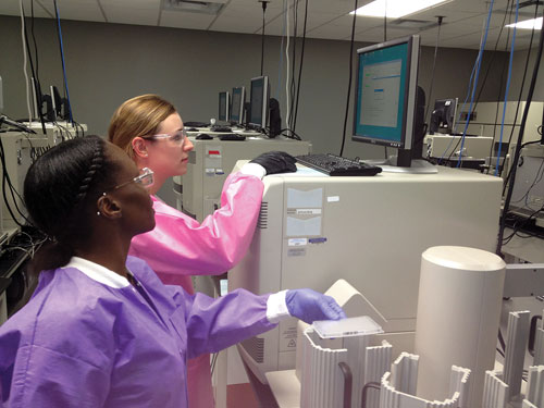
A graduate student at UNMC analyzes a Western blot from an immunodepletion assay that was used to examine the regulation of Aurora B kinase activity by TPX2. [Jintana Saowapa]
Discovery and Development
Beyond fundamental research studies, scientists in both academic and industrial labs are looking to kinase assays to discover and develop new anticancer treatments.
Oncolytic viruses are a promising approach to treating cancer due to their potential to selectively kill cancer cells in the midst of healthy tissue. In a collaboration spearheaded by Bruce Bejcek, Ph.D., and Karim Essani, Ph.D., both professors at Western Michigan University, researchers are currently exploring the potential of tanopox virus (TPV) as an additional tool in the arsenal of oncolytic therapies.
Tumor suppressor protein p53 is a promising target for virotherapy since it is mutated in more than 50% of cancers. To determine the potential of TPV as an oncolytic therapy, Dr. Bejcek’s team performed in vitro assays with the 142R protein of TPV, which has a serine-threonine kinase domain that is homologous to the p53-phosphorylating B1R gene in the more well-known vaccinia virus. The team found that 142R interacts with p53 in vitro and is thus a good candidate for creating a conditionally replicating virus that can selectively target cancer cells and leave healthy cells unharmed.
The kinase assay involved mixing recombinant, bacterially expressed 142R and p53 in the presence of radiolabeled ATP. The researchers ran the proteins out on a gel and observed that radioactive phosphate was added onto p53 only in the presence of 142R, suggesting that 142R phosphorylates p53. The team plans to determine if 142R is able to phosphorylate p53 in cells, and also hope to determine whether knocking out 142R can cause TPV viruses to selectively replicate in, and ultimately kill, cancer cells lacking wild-type p53, Dr. Bejcek says.
While kinase assay platforms that probe interactions between inhibitors and a single kinase are sufficient for certain applications, for others, researchers require methods to rapidly and quantitatively characterize hundreds of kinases. With this end in mind, researchers at Ambit Biosciences and DiscoveRx developed a high-throughput method for yielding quantitative ligand-binding data for kinase inhibitors across greater than 80% of the human kinome, which contains more than 500 members.
Once a kinase inhibitor is discovered in initial screening assays, researchers need to determine how those inhibitors interact across the broader kinome, says Daniel Treiber, senior director of research and development at DiscoveRx. A team of Ambit and DiscoveRx scientists, led by Treiber and Patrick Zarrinkar (now at Blueprint Medicines), developed a method known as KINOMEscan and have used this technology to screen the interactions of 72 known, mature kinase inhibitors across 442 kinases in vitro.
KINOMEscan is based on a competitive binding assay that combines affinity chromatography and ultrasensitive protein detection. Researchers immobilize a known kinase inhibitor on a solid support and then introduce a kinase and determine how much kinase is captured in the presence or absence of a test inhibitor.
The kinases, which are labeled with a DNA strand, are then eluted off the solid support, and researchers perform qPCR to quantify the amount of bound kinase. In this way, they determine how effective the test inhibitors are at inhibiting the kinase-ligand interaction and are able to produce full dose-response curves that yield thermodynamic binding constant (Kd) values.
The DiscoveRx team, working closely with epigenetics-based drug discovery pioneer James Bradner of the Dana Farber Cancer Institute, has taken the KINOMEscan technology and extended it to study inhibitors of bromodomains, a class of protein domains that selectively bind to acetylated histone tails and are thought to be disease drivers.
Whether each of the 56 known bromodomains is druggable and can be selectively targeted with an inhibitor remains to be seen, says Treiber, who led a team of DiscoveRx scientists to develop an adapted form of the KINOMEscan methodology to screen broadly for bromodomain inhibitors. So far, Treiber’s team has validated the technology for about 20% of bromodomains and their findings are consistent with and validated by previous literature reports.
When developing an assay to screen for protein inhibitors, researchers often have to compromise quantitative rigor and the number of proteins screened simultaneously to achieve high throughput. According to Treiber, the beauty of the KINOMEscan approach is it has all of these qualities, thanks to the ultrasensitive and quantitative nature of qPCR and the unique semi-automated and generic high-throughput setup.
“The assays are exquisitely sensitive, so it allows us to play games that you can’t play with fluorescence or other traditional technologies,” Treiber says.
Up next, the research team plans to expand upon the bromodomain panel, and then begin to offer it as a service to customers interested in drug screening and optimization.
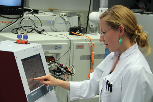
Scientists at DiscoveRx working with KINOMEscan prepare to analyze samples on an ABI 7900 qPCR instrument. [J. Hunt]
Characterization of Kinase Inhibitors
Once a drug candidate has been identified and validated in initial screens, researchers need to characterize its effects in cancer cells before progressing into clinical trials. One way to determine if a kinase inhibitor is effective in cancer cells is to determine whether treatment with the inhibitor reduces the phosphorylation of proteins that are downstream of the targeted kinase.
Btk is a known regulator of B-cell receptors and is implicated in malignant B-cell survival. Researchers at Ono Pharmaceuticals wanted to find out how ONO-WG-307, a known Btk kinase inhibitor, affected Btk-dependent signal transduction in two tumor cell lines. In a study performed for Ono Pharmaceuticals, researchers, led by Henrik Daub, Ph.D., svp of science and technology at Evotec, used LC/MS to perform a quantitative phosphoproteomic analysis to help shed light on the cellular mode of action of ONO-WG-307.
The general approach involves incubating cancer cells with isotopically labeled amino acids, which introduces heavier-weight variants into the proteome and enables quantitation, Dr. Daub says. Researchers then incubate the cancer cells in the presence or absence of the compound, lyse the cells, and cleave cellular proteins with a protease.
To isolate the peptide fraction from this extremely complex mixture, researchers use chromatography and then further enrich for phosphorylated peptides. Finally, researchers analyze the enriched phosphopeptides on a LTQ-Orbitrap-Velos mass spectrometer and perform bioinformatic processing to analyze the data. Any changes observed in the intensities of the peptide signals from drug-treated cells, compared to control cells, suggest a change in cellular activity as a result of the drug treatment.
“We typically get a lot of data,” Dr. Daub says, “on the order of up to several hundred thousand peptide identifications initially.” After removing redundant peptide signals, the result is about 10,000 or 15,000 quantifiable signals from distinct phosphorylation sites. Such analyses can yield a wealth of useful information about which downstream signaling pathways are affected by a drug.
In the future, Dr. Daub hopes to see this quantitative phosphoproteomics approach taken to a new level of biological analysis.
“There are quite some interesting developments which can be anticipated,” Dr. Daub explains. For example, Dr. Daub says he hopes to be able to determine not only which phosporylation changes are occurring, but also where in the cell these changes are taking place.
This should be achievable by collecting and analyzing subcellular fractions, Dr. Daub insists. Such analyses done over a time course could also reveal if the process of signaling changes the subcellular location of the proteins.
In addition to shedding light on the cellular mode of action of a kinase inhibitor, quantitative phosphoproteomic analyses can also reveal why a compound may have effects in certain cancer cell lines and not others, and help researchers determine if a promising drug candidate has any off-target effects.
“The strength of doing this in a very broad or more or less global manner is that it’s unbiased,” Daub remarks.
Knowing how a compound works can be just as important in determining that it does work. Quantitative phosphoproteomics makes it possible for researchers “to identify unexpected new things…and get some interesting hints and insights about how and why the kinase inhibitor exerts its therapeutic effects,” Dr. Daub says.
Researchers at Evotec used LC/MS to perform a quantitative phosphoproteomic analysis to help shed light on the cellular mode of action of Ono Pharmaceuticals’ ONO-WG-307. [Dirk Ullmann]

