July 1, 2016 (Vol. 36, No. 13)
Hanging-Drop Technology Permits Scaffoldless Modeling of Drug-Induced Liver Injury
InSphero, a developer of three-dimensional (3D) cell culture technology, offers microtissue platforms derived from liver, pancreas, tumor, heart, brain, and skin. These platforms constitute the company’s 3D Insight™ line, which was expanded recently to include what the company calls Multi-donor Human Liver Microtissues. The new platform incorporates pooled hepatocytes from multiple donors and offers a more predictive model for drug-induced liver injury (DILI).
The new platform, like each of the other 3D Insight platforms, supports 3D spheroids while eschewing scaffolds. According to InSphero, a scaffold-free approach is attractive because it allows microtissue formation to be driven by completely native cell-cell interactions. Also, the absence of scaffold material facilitates a greater number of downstream assays and imaging options.
The new platform is the logical extension of InSphero’s core philosophy of creating assay-ready tissues for the life sciences industry. By incorporating hepatocytes from five male and five female donors, the new platform manifests genetic diversity, a quality that enables researchers to study the effects of compounds of interest on the general population, distinguishing between known hepatotoxicants and related nontoxic analogs.

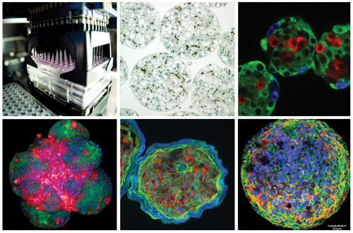
InSphero aims to improve in vitro testing by facilitating the development and use of organotypic 3D cell culture models. Top left image: InSphero’s patented GravityPLUS™ Hanging Drop System. Subsequent images (in clockwise order): assay-ready 3D liver, islet, tumor, skin, and brain microtissues. The GravityPLUS can manufacture all these microtissues for safety and efficacy testing.
Benefits of Assay-Ready Microtissue
For cell-based assays to be more efficient and biologically relevant, it helps for researchers to use standardized 3D models,” asserts Jan Lichtenberg, Ph.D., InSphero’s CEO and co-founder. Standardization enables scientists to conduct benchmark studies that are meaningful to other researchers without the error-inducing need to convert home brews to standard references. “Optimizing cell-based knowledge,” adds Dr. Lichtenberg, “yields a cost-to-benefit ratio that enables researchers everywhere to generate data that can be published and easily compared.”
3D Hanging Drop Culture
Many types of cells form the tissues that make up the liver, heart, and other structures in the body. To understand drug interactions with those tissues, scientists need to assay compounds not just in a single cell type, but in models that mimic tissue in vivo.
The 2D monolayer and traditional 3D scaffolding techniques used to create tissue models, however, have limitations that affect their value to scientists. For example, 2D monolayers fail to replicate gradients in nutrients, oxygenation, metabolites, and proliferation. Another issue is waning functionality. “Hepatic cells grown in a 2D environment lose functionality in two to four days,” observes Dr. Lichtenberg. “Therefore, any toxic effects that appear 7 to 10 days after treatment are beyond the scope of what is seen in the lab.”
With 3D scaffolding techniques, “spheroid sizes may vary, and matrix materials may interfere with downstream analyses. “The hydrogels and other scaffolding materials introduce artificial material into the cell tissue,” explains Dr. Lichtenberg. “Many scaffolds are porous, making tissue less dense and allowing drug molecules to stick to the scaffold, which makes them less available to the tissue.”
To overcome these challenges and add scalability, InSphero uses ultra-low attachment (ULA) and hanging-drop production technologies. For example, the GravityPLUS™ platform, which exploits the hanging-drop approach, relies on gravity to form 3D structures within automation-compatible 96-well plates. “With GravityPLUS,” says Dr. Lichtenberg, “we create a dense, tissue-like structure that is morphologically and functionally very similar to native tissue.” InSphero’s hepatic tissues, for instance, are still functional after five weeks. Lab reports suggest that functionality continues for 60 to 70 days.
Broad Applications
InSphero’s 3D InSight microtissues are based on the company’s proprietary cell selection and preconditioning techniques. At present, InSphero offers models for the liver, pancreas, tumor, heart, brain, and skin. “But we can form tissues with nearly every cell type in the body,” insists Dr. Lichtenberg. “Ours is a true umbrella patent.”
The GravityPLUS platform lets scientists use a small number of cells to produce a lot of data points at a price that allows easy technological integration into their workflow. Once researchers integrate the microtissues into their work and deploy the requisite downstream processes, they can scale operations easily. “It’s a matter of ordering product, taking off the lid, and starting testing,” states Dr. Lichtenberg.
That accessibility and relatively low cost encourages the use of microtissues not only during late-stage work, but also during early-stage discovery and validation of potential lead candidates, Dr. Lichtenberg tells GEN.
Growth and Development
InSphero’s position as a privately held company allows it the freedom to innovate without the pressures of producing quarterly reports for shareholders. As Dr. Lichtenberg recounts, “When we worked on our financing round last year, we considered our options and opportunities to capitalize the company. We decided that being a private company had helped us develop quickly during the past few years.”
“We’re at an exciting stage,” Dr. Lichtenberg continues. “We see the adoption of 3D microtissue technology globally, and we have extended our sales force and customer support in the United States and worldwide.” InSphero also recently completed a new production facility in the United States., enabling product to reach American customers faster.
The company is also focusing on developing integrated solutions and enhancing compatibility. “We’re interested in offering a full solution for our customers,” comments Dr. Lichtenberg. To that end, InSphero works closely with assay and imaging technology providers and is considering possible acquisitions.
Microtissues for Diagnostics
The company also is expanding its pipeline to include disease models though its new division, InSphero Diagnostics. It has used its technology to generate microtissues that are based on patient biopsies and that mimic patients’ tumors. Drugs can be tested against such microtissues to determine which intervention is most successful for a given patient. “We’re in early-stage clinical feasibility studies now,” says Dr. Lichtenberg. He anticipates that commercialization will begin in a couple of years.
At InSphero, diagnostic work and predictive work are symbiotic. “We make our models more relevant to physicians and researchers by moving models from the cell lines typically used for screening to more specific cell types—PDX or even primary tumor cells—that result in more relevant tests,” details Dr. Lichtenberg. “We’re also combining microtissues to mimic interactions between tissues. This will lead to body-on-a-chip applications, which we are developing in cooperation with AstraZeneca in the U.K.”
The body-on-a-chip work was one of the largest publically funded projects in Europe on microphysiological systems, involving six partners, he notes. Data was published last year in Nature Communications. “We’ve started beta tests and plan to make the technology available to more beta partners in 2017,” informs Dr. Lichtenberg. “We have some clear applications in mind, including a kit to solve a particular drug-testing problem.”
InSphero
Location: Wagistrasse 27, CH-8952 Schlieren, Switzerland
Phone: +41 44 515 04 90
Website: www.insphero.com
Principal: Jan Lichtenberg, Ph.D., CEO and Co-founder
Number of Employees: 70
Focus: InSphero develops scalable, assay-ready 3D microtissues using its patented hanging-drop technology, which eliminates the need for scaffolds.


