Scientists at Brown University have developed a method for creating 3-D-printed biomaterials that can degrade on demand. The team says their technique can be used to help make microfluidic devices or cell cultures than can change during experiments.
“It's a bit like Legos,” notes Ian Wong, Ph.D., an assistant professor in Brown's School of Engineering and co-author of the research. “We can attach polymers together to build 3-D structures, and then gently detach them again under biocompatible conditions.”
The study (“Stereolithographic Printing of Ionically-Crosslinked Alginate Hydrogels for Degradable Biomaterials and Microfluidics”) is published in Lab on a Chip.
“…we show stereolithographic printing of hydrogels using noncovalent (ionic) crosslinking…enables reversible patterning with controlled degradation,” write the investigators. “We demonstrate this approach using sodium alginate, photoacid generators and various combinations of divalent cation salts, which can be used to tune the hydrogel degradation kinetics, pattern fidelity, and mechanical properties.”
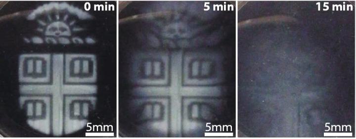
![Brown researchers have found a way to 3-D print intricate temporary microstructures that can be degraded on demand using a biocompatible chemical trigger. The technique could be useful could be useful in fabricating microfluidic devices, creating biomaterials that respond dynamically to stimuli and in patterning artificial tissue. [Wong Lab / Brown University]](https://genengnews.com/wp-content/uploads/2018/08/Sep8_2017_BrownUniv_3DPrintMicrostructures1695672166-1.jpg)
Brown researchers have found a way to 3-D print intricate temporary microstructures that can be degraded on demand using a biocompatible chemical trigger. The technique could be useful could be useful in fabricating microfluidic devices, creating biomaterials that respond dynamically to stimuli and in patterning artificial tissue. [Wong Lab / Brown University]
Stereolithography uses an ultraviolet laser controlled by a computer-aided design system to trace patterns across the surface of a photoactive polymer solution. The light causes the polymers to link together, forming solid 3-D structures from the solution. The tracing process is repeated until an entire object is built from the bottom up.
Stereolithographic printing ordinarily involves photoactive polymers that link together with irreversible covalent bonds. Dr. Wong and colleagues wanted to make structures with potentially reversible ionic bonds, which had never been done before using light-based 3-D printing. To do so, researchers made precursor solutions with sodium alginate, which is obtained from seaweed that can ionically crosslink.
“The idea is that the attachments between polymers should come apart when the ions are removed, which we can do by adding a chelating agent that grabs all the ions,” continues Dr. Wong. “This way we can pattern transient structures that dissolve away when we want them to.”
The researchers showed that alginate could be used in stereolithography. With different combinations of ionic salts (magnesium, barium, and calcium) they made structures with varying stiffness, which could then be dissolved at varying rates.
“It's a helpful tool for fabrication,” says Thomas M. Valentin, a Ph.D. student in Wong's lab and the study's lead author. For example, the scientists demonstrated that they could use alginate as a template for making lab-on-a-chip devices with microfluidic channels.
“We can print the shape of the channel using alginate, then print a permanent structure around it using a second biomaterial,” points out Valentin. “Then we simply dissolve away the alginate and we have a hollow channel. We don't have to do any cutting or complex assembly.”
The team also demonstrated that degradable alginate structures can create dynamic environments for experiments with live cells. They carried out a number of studies with alginate barriers surrounded by human mammary cells and looked at how the cells migrate when the barrier is dissolved. These kinds of experiments can be useful in investigating wound-healing processes or the migration of cells in cancer, according to the researchers, who add that neither the alginate barrier nor the chelating agent used to dissolve it away caused any serious toxicity to the cells.
The biocompatibility of the alginate is promising for additional future applications, including in making scaffolds for artificial tissue and organs, notes Dr. Wong.
“We can start to think about using this in artificial tissues where you might want channels running through it that mimic blood vessels,” he says.


