
Engineers at Rutgers used 4D printing to create tiny needles that mimic parasites that attach to tissues and could replace hypodermic needles. The team published its study (“4D Printing of a Bioinspired Microneedle Array with Backward‐Facing Barbs for Enhanced Tissue Adhesion”) in Advanced Functional Materials.
While 3D printing builds objects layer by layer, 4D goes further with smart materials that are programmed to change shape after printing. Time is the fourth dimension that allows materials to morph into new shapes.
“Microneedle (MN), a miniaturized needle with a length‐scale of hundreds of micrometers, has received a great deal of attention because of its minimally invasive, pain‐free, and easy‐to‐use nature. However, a major challenge for controlled long‐term drug delivery or biosensing using MN is its low tissue adhesion. Although microscopic structures with high tissue adhesion are found from living creatures in nature (e.g., microhooks of parasites, barbed stingers of honeybees, quills of porcupines), creating MNs with such complex microscopic features is still challenging with traditional fabrication methods,” write the investigators.
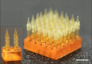
“Here, a MN with bioinspired backward‐facing curved barbs for enhanced tissue adhesion, manufactured by a digital light processing 3D printing technique, is presented. Backward‐facing barbs on a MN are created by desolvation‐induced deformation utilizing cross‐linking density gradient in a photocurable polymer. Barb thickness and bending curvature are controlled by printing parameters and material composition. It is demonstrated that tissue adhesion of a backward‐facing barbed MN is 18 times stronger than that of barbless MN. Also demonstrated is sustained drug release with barbed MNs in tissue. Improved tissue adhesion of the bioinspired MN allows for more stable and robust performance for drug delivery, biofluid collection, and biosensing.”
“We think our 4D-printed microneedle array will allow for more robust and sustained use of minimally invasive, pain-free and easy-to-use microneedles for delivering drugs, healing wounds, biosensing and other soft tissue applications,” said senior author Howon Lee, PhD, an assistant professor in the department in the School of Engineering at Rutgers University-New Brunswick.
This YouTube video by Riddish Morde shows microneedles applied to chicken muscle tissue.
Hypodermic needles are widely used in hospitals and labs to extract blood and inject drugs, causing pain, scarring skin and posing an infection risk. People with diabetes often take blood samples multiple times a day with needles to monitor blood sugar levels.
Microneedles are gaining attention because they are short, thin and minimally invasive, reduce pain and the risk of infection and are easy-to-use. But their weak adhesion to tissues is a major challenge for controlled drug delivery over the long run or for biosensing, which involves using a device to detect DNA, enzymes, antibodies and other health indicators. Inspired by the living creatures in nature that have developed microscopic features that adhere to tissue, Rutgers engineers developed a microneedle that interlocks with tissue when inserted, enhancing adhesion. They combined a micro 3D-printing technique and a 4D-printing approach to create backward-facing barbs on a microneedle.
Using chicken muscle tissue as a model, the researchers showed that tissue adhesion with their microneedle is 18 times stronger than with a barbless microneedle. Their creation outperforms previously reported examples, resulting in more stable and robust drug delivery, collection of bio-fluids and biosensing, the study says.


