
Source: Allen Institute
An international team of scientists headed by researchers at the Allen Institute for Brain Science has generated what may be the most detailed “parts list” of the human brain to date. Their research, described in Nature, mapped the organization of individual cell types in different layers of the human cortex, and produced an ultra-high resolution comparison between the human and mouse brain cell types. The results indicated that that the majority of human brain cells have a mouse equivalent that has effectively been retained down 75 million years of evolution. However, there were also extensive differences between the homologous human and mouse cell types, and the researchers suggest that key differences between human and rodent brain structure and cell organization could help to explain why so many psychiatric drugs developed using mouse models don’t work in humans.
“The bottom line is there are great similarities and differences between our brain and that of the mouse,” said Christof Koch, PhD, chief scientist and president of the Allen Institute for Brain Science. “One of these tells us that there is great evolutionary continuity, and the other tells us that we are unique. If you want to cure human brain diseases, you have to understand the uniqueness of the human brain.” Koch and colleagues describe their findings in a paper titled “Conserved cell types with divergent features in human versus mouse cortex.”
Source: Allen Institute
The human cerebral cortex processes our higher cognitive abilities and functions, and is the “most complex structure known to biology,” the authors wrote. It is made up of 16 billion neurons and 61 billion non-neuronal cells, which are organized into more than 100 distinct anatomical or functional regions. When compared with the cortex of the mouse, which is the animal model most commonly used for research, the human cortex has a more than 1,000-fold larger area and number of neurons.
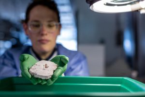
The researchers used a technique called single-nucleus RNA-sequencing (snRNA-seq) to profile the gene expression of individual nuclei from frozen human brain specimens, and identify the different types of neurons and non-neuronal brain cell in different layers of an area of the human cortex called the middle temporal gyrus (MTG). “In total, 15,928 nuclei passed quality control, including those from 10,708 excitatory neurons, 4,297 inhibitory neurons and 923 non-neuronal cells,” they noted.
The cell analyses identified 6 non-neuronal, 24 excitatory, and 45 inhibitory cell types in the human MTG. A similar Allen Institute study in mice, published last year, provided a dataset of mouse cells, which the team then used for comparison. This showed that the 75 human brain cell types have basic equivalents in the mouse, so while the brains if humans and mice may look different, at the gene expression level, the cell types are pretty similar.
“Beyond similarities in overall diversity and hierarchical organization, most cell types mapped at the subclass level, seven cell types mapped one-to-one, and no major classes had missing homologous types despite the last common ancestor between humans and mice living at least 65 million years ago, and despite the thousand-fold difference in brain size and number of cells,” the authors reported. “Therefore, the transcriptomic organization of cell classes and subclasses appears conserved, with species and regional variation found at the finest level of cell-type distinction.”
There were many differences between the two species, however. Serotonin receptors, which are found on neurons that react to the neurotransmitter serotonin, and which are key to appetite, mood, memory, sleep and many other important brain functions, are hugely different between mouse and human brain cells, the study found. While humans and mice both have these proteins, they are not found in the same kinds of neurons in the two species. This key difference could explain why it’s been so difficult to develop new antidepressants, or drugs for other serotonin-related disorders. A drug that acts on a serotonin receptor could have very different effects in mice and in humans. “… these results help resolve the paradox of failures in the use of mouse for preclinical studies despite conserved structure across mammals, and highlight the need to analyse human brain in addition to model organisms,” the team stated.
“Many people would assume that the human brain is more complex than the mouse brain,” said Ed Lein, PhD, investigator at the Allen Institute for Brain Science, and senior author on the study. “It’s somewhat of a surprise that at least in terms of cellular diversity, that doesn’t seem to be the case. But now that we have a way to make a true apples-to-apples comparison, we start to see many differences in how the genes are used, what the cells look like and the proportions of different cell types in the brain.”
It’s hoped that the new comparison of and alignment between mouse and human brain cell types will help scientists extrapolate knowledge acquired over decades of rodent research to the human brain. “The parts list of the human brain is key,” said Joshua Gordon, MD, PhD, director of the National Institute of Mental Health. “That list allows us to understand what’s different between mice and human beings and what’s similar. The ultimate impact of this understanding will be better treatments for mental illnesses. The eventual goal will be to develop the complete parts list of the entire human brain. It’s a hugely ambitious goal, but this paper shows us that it’s a doable undertaking.”


