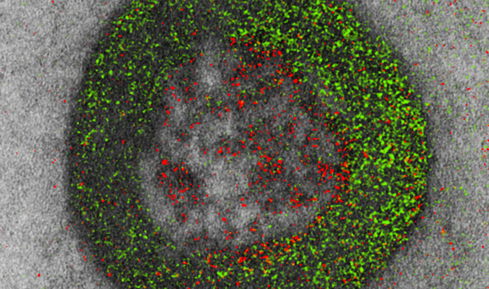
G protein–coupled receptors (GPCRs) initiate signaling cascades while ensconced in the cell membrane. At least, that’s where GPCRs are usually found. But some GPCRs ride endosomes into the cell’s interior. What’s more, these roving GPCRs add a spatial dimension to signaling, report scientists based at the University of California, San Diego (UCSD). Indeed, in a recent Nature article, the scientists suggested that intracellular, endosome-embedded GPCRs contribute to “diverse GPCR biology, cancerous dysregulation, and therapeutic vulnerability.”
The new information about GPCRs appeared in an article titled, “Non-canonical β-adrenergic activation of ERK at endosomes.” The article’s senior author, Jin Zhang, PhD, asserted that her group’s findings “may change the textbook model of GPCR-mediated signaling, and that could have profound implications for future drug development.”
“Here we investigated how spatially organized β2-adrenergic receptor (β2AR) signaling controls ERK,” the article indicated. “Using subcellularly targeted ERK activity biosensors, we show that β2AR signaling induces ERK activity at endosomes, but not at the plasma membrane.”
The enzyme called extracellular-signal-regulated kinase (ERK) regulates transcription of a number of genes involved in controlling cell growth. When ERK signaling goes awry, several pathologies, including cancer, may arise. As such, therapeutic intervention targeting members of the ERK signaling pathway is a major endeavor among cancer researchers.
“[The endosomal] pool of ERK activity depends on active, endosome-localized Gαs and requires ligand-stimulated β2AR endocytosis,” the article’s authors continued. “We further identify an endosomally localized noncanonical signaling axis comprising Gαs, RAF, and mitogen-activated protein kinase, resulting in endosomal ERK activity that propagates into the nucleus. Selective inhibition of endosomal β2AR and Gαs signaling blunted nuclear ERK activity, MYC gene expression, and cell proliferation.”
These findings help clarify the mechanism underlying GPCR-regulated ERK activation. This mechanism has long been a mystery.
“We found that GPCR-mediated ERK signaling, upon activation at the endosomes, propagates into the nucleus, where it turns on important genes to control cell growth,” Zhang explained. “Given the closer proximity between endosomes and the nucleus, compared with the plasma membrane and nucleus, perhaps cells take advantage of the 3D spatial organization of cellular organelles and use a ‘short cut’ for efficient receptor signal transduction.”
According to textbook models, GPCRs can signal through two distinct classes of molecules: G proteins and arrestins. The new research suggests greater diversity in how GPCRs signal downstream.
Arrestins, intracellular proteins involved in terminating plasma membrane signaling, were thought to serve as a scaffold to enable ERK activation. “Our data strongly support arrestin’s involvement in ERK activation, but through its capacity to help internalize the receptor and not as a scaffold for ERK as previously thought,” said co-author Roger Sunahara, PhD, professor of pharmacology, UCSD.
Zhang added that the new work suggests arrestins and G proteins work collaboratively to activate ERK at the endosomes, with arrestins escorting the receptors to endosomes and G proteins recruiting the ERK activation machinery.
“There are broad implications for both basic and translational science considering the large number of GPCRs involved in passing on various cellular messages to regulate body functions,” Zhang noted. “An immediate implication is the potential generality of the proposed model, which should be investigated beyond the few receptors we studied in this work.
“In terms of translational impact, GPCR drug development has been influenced by concepts like ‘biased signaling,’ with drugs developed to preferentially activate G proteins or arrestins. The discovery that some receptors require the collaboration of arrestins and G proteins to activate ERK is expected to change the way GPCR drugs are developed.”
Many types of cancers contain mutations in G proteins that contribute to cancer development, said UCSD’s J. Silvio Gutkind, PhD. He was not one of the current paper’s authors, but he offered this observation: “Many tumors harbor persistently active G proteins and GPCRs, including cancers of the colon, pancreas, and appendix. The new findings can now be exploited to develop new treatment strategies to prevent and treat these human malignancies.”


