![Systems microscopy and colorectal cancer projects will be based at Karolinska’s Center for Biosciences.[rgbspace-Fotolia.com]](https://www.genengnews.com/wp-content/uploads/2018/08/UGENWebsitepicturesTopStoriesFeb2011Feb10_2011_4595195_EnlargedDNAunderMicroscope_EUfundingforTwoCancerConsortia2341312011.jpg "Systems microscopy and colorectal cancer projects will be based at Karolinska’s Center for Biosciences.[rgbspace-Fotolia.com]")
Unlike other modes of microscopy that employ sophisticated optical equipment to acquire high-resolution images, expansion microscopy enlarges biological specimens physically enabling nanoscale image acquisition using conventional microscopes. The technology is based on hydrogel polymers that swell with water and bind biomolecules to homogenously expand tissue samples. One of the drawbacks, however, of current expansion microscopy protocols is that sample preparation is a multistep process that involves pre-treatment with anchoring chemicals that link different labels and biomolecules to the gel.
In a paper published in the journal Nature Biotechnology on January 2, 2023, “Magnify is a universal molecular anchoring strategy for expansion microscopy,” a team of scientists from Carnegie Mellon University, the University of Pittsburgh and Brown University report a new method called ‘Magnify’ that uses a novel, mechanically sturdy hydrogel that holds different classes of biomolecules such as nucleic acids, proteins and lipids, without the need for additional steps to anchor these molecules to the gel.
The new technology improves the convenience and applicability of expansion microscopy and could benefit a variety of biomedical investigations that depend on visual data.
“Magnify can be a potent and accessible tool for the biotechnology community,” said Yongxin (Leon) Zhao, PhD, an associate professor of biological sciences at Carnegie Mellon University and the senior author of the study. “We overcame some of the longstanding challenges of expansion microscopy. One of the main selling points for Magnify is the universal strategy to keep the tissue’s biomolecules, including proteins, nucleus snippets and carbohydrates, within the expanded sample.”
Aleksandra Klimas, PhD, a postdoctoral researcher, and Brendan Gallagher, a graduate student, were first co-authors on the paper. “This is an accessible way to image specimens in high resolution,” said Klimas. “Traditionally, you need expensive equipment and specific reagents and training. This method is broadly applicable to many types of sample preparations and can be viewed with standard microscopes that you would have in a biology laboratory.”
A problem of expansion
Magnify’s new hydrogel formula, invented in Zhao’s lab, expands biological samples up to 11 times, enabling imaging of a variety of cells and tissues. The new hydrogel is more robust than its predecessor, which was very fragile, causing breaks during the imaging process.
Earlier methods adopted to expand cells for expansion microscopy used enzymes to digest proteins (proteases). Therefore, the labelled gel was essentially a surrogate for the location previously occupied by a protein. The new method not only keeps proteins and other biomolecules intact, it allows the detection of different types of biomolecules in the same sample, including proteins, lipids, carbohydrates, and nucleic acids.
“One of the key concepts that we tried to keep in mind was to meet researchers where they are and have them change as few things in their protocols as possible,” said Gallagher. “It works with different tissue types, fixation methods and even tissue that has been preserved and stored. It is very flexible. You don’t necessarily need to redesign experiments with Magnify in mind completely. It will work with what you have already.”
Resolution and versatility
The ability of conventional optical microscopes or ‘light microscopes’ (that use visible light and a system of lenses to magnify images of samples) to distinguish two points (resolution) in a sample is limited by the natural scattering of light when it hits an object (diffraction). This limits the resolution of light microscopes to around 0.2 micrometer (200 nanometer).
When used with a diffraction-limited objective lens on conventional optical microscopes, Magnify offers a much higher resolution of about 25 nanometers, and when combined with super-resolution optical fluctuation imaging, the new technique offers a resolution of nearly 15 nanometers.
Broad applications
Co-author of the study, Simon Watkins, PhD, founder and director of the Center for Biologic Imaging at the University of Pittsburgh and the Pittsburgh Cancer Institute said, “Let’s say you have a tissue with dense and non-dense components, this gets around tissues that previously wouldn’t expand isometrically. Leon has been working hard on this to make this protocol work with tissues that have been archived.”
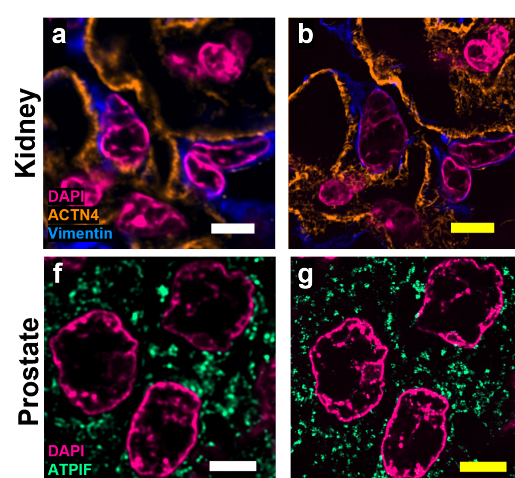
The authors demonstrate Magnify can be applied to visualize a broad range of tissue types at nanoscale resolution including proteins in the synapses of mouse brains, podocytes or foot processes in kidney cells of formalin-fixed and paraffin-embedded human tissue, and individual ciliary apparatus in human lung organoids.
Working in collaboration with Zhao’s lab, Xi (Charlie) Ren, PhD, an assistant professor of biomedical engineering at Carnegie Mellon, developed lung organoid models with structural defects in cilia to validate the ability of Magnify to visualize clinically relevant ciliary pathology. “With the latest Magnify techniques, we can expand lung tissues and start to see some ultrastructure of the motile cilia even with a regular microscope. This will expedite both basic and clinical investigations,” said Ren.
Future steps
Co-author Alison Barth, PhD, a professor in the life sciences at Carnegie Mellon said the broad applications provided by the new method will be a boon for researchers. “The brain is a great place to take advantage of these super-resolution techniques,” said Barth. “Microscopy methods will be beneficial for synaptic phenotyping and analysis across different brain conditions”
Zhao and his team of collaborators hope to develop the Magnify technology further to increase its accessibility among basic and applied researchers.
This work was funded by Carnegie Mellon, the Kaufman Foundation, the DSF Charitable Foundation, the US Department of Defense, the National Institutes of Health (NIH), the Air Force Office of Scientific Research, NeuroNex and Brown University.


