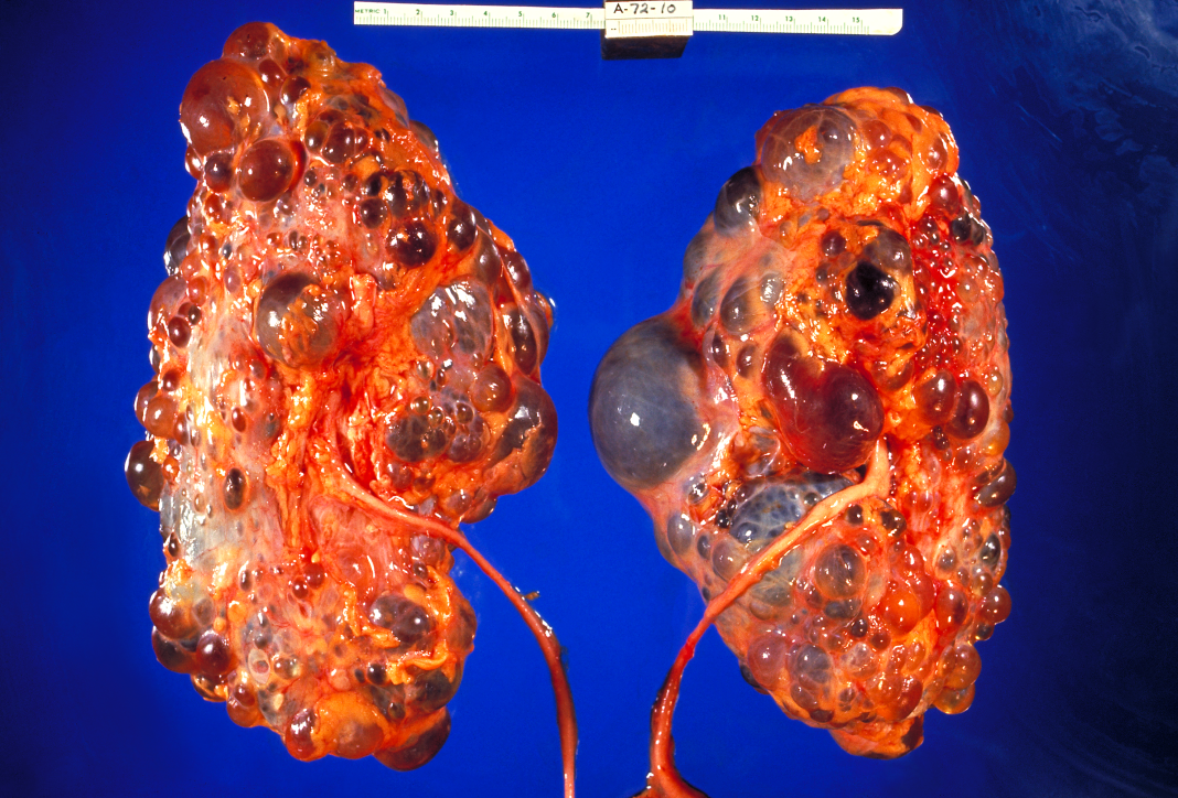
A new study published in the Journal of the American Society of Nephrology underscores the importance of the channel protein polycystin-2 in the endoplasmic reticulum (ER) in the maintenance of kidney health, and provides evidence that dysfunction of the gene in the ER can lead to the common and currently incurable condition called autosomal dominant polycystic kidney disease (ADPKD), where the growth of cysts lead to kidney failure.
The new findings published in the article titled, “Channel Function of Polycystin-2 in the Endoplasmic Reticulum Protects against Autosomal Dominant Polycystic Kidney Disease,” overturn the prevailing view that polycystin-2 functions as a calcium channel in a key cellular appendage called the primary cilium to prevent the growth of cysts in the kidney. This view classifies ADPKD as a “disease of the primary cilia” or a “ciliopathy.” The insights of the current study uncover an overlooked mechanism that could lead to new strategies to treat ADPKD.

Senior author of the study Chou-Long Huang, MD, PhD, a professor at the University of Iowa Carver College of Medicine, said, “ADPKD is a very common genetic kidney disease and accounts for 5–10% of ESRD [end stage renal disease] patients. There is no effective treatment thus far. Disease mechanism remains largely unknown, preventing development of effective therapy.”
Mutations in the genes encoding polycystin-1 and -2 cause most cases of ADPKD. Recent studies have revealed polycystin-2 is primarily localized to the ER and acts more as a gateway for potassium than calcium ions. Until now it has been unclear whether polycystin-1 and -2 in cellular locations other than the primary cilia play a role in ADPKD.
To answer this question, Huang and his colleagues examined the role of polycystin-2 in the ER, a labyrinth of interconnected membranous tunnels in the cell’s cytoplasm involved in protein and lipid synthesis. The researchers revealed polycystin-2 in the ER is important for kidney health and that its loss in the ER can lead to cyst formation.
A potassium channel in the ER membrane called TRIC-B (trimeric intracellular cation channel-B) exchanges potassium and calcium ions, releasing calcium ions into the cytosol. “Using TRIC-B as a tool, we examined the function of ER-localized polycystin-2 and its role in ADPKD pathogenesis in cultured cells, zebrafish, and mouse models,” the authors noted.
When the researchers bred mice lacking polycystin-2, the researchers found the release of calcium ions from the ER was defective in cells lacking polycystin-2 and the animals developed cysts. The authors then exogenously expressed TRIC-B in the mice lacking polycystin-2 in the ER and found this prevented the growth of cysts. On the other hand, TRIC-B deletion further exacerbated the growth of cysts in mice missing one allele of the polycystin-2 gene in the kidney.
“The study challenges the current dogma that the disease mechanism of ADPKD is predominantly due to defects in the cilia. We show that function of polycystin-2 in the ER plays a crucial role in preventing cyst formation in ADPKD,” said Huang. “Moreover, polycystin-2 functions as a potassium ion channel rather than a calcium ion channel as is currently believed. The study suggests that direct activation of polycystin-2 function in the ER will be an effective treatment for ADPKD.”
The new data also indicates that activating TRIC-B in AKPD patients could be an effective pharmacotherapeutic strategy.
In future studies, Huang’s team intends to investigate whether activation of polycystin-2 function in the ER can prevent ADPKD caused by mutations in polycystin-1 and reverse the disease after cysts have already formed in the kidney.


