
Stanford University researchers developed a tissue-mimicking, stretchable sensor termed NeuroString for real-time measurement of neurotransmitter dynamics in vivo in the brain, gut, and other soft organs. The elastic and conformable biosensing interface has broad potential for studying the impact of neurotransmitters on brain-gut communication and may be extended to biomolecular sensing in other soft organs across the body.
The soft biomolecular monitor is described in Nature in an article titled, “A tissue-like neurotransmitter sensor for the brain and gut.”
Neurotransmitters—signaling molecules produced and released by neurons—play essential roles in regulating neural circuit dynamics in the central nervous system (CNS) and the peripheral, including the gastrointestinal tract.
In the CNS, monoamines, including dopamine (DA) and serotonin (5-HT), are involved in the regulation of cognitive processes such as emotion, arousal, and memory. Dysregulated monoamine signaling is a common feature of many psychiatric and neurological disorders, including addiction, major depressive disorder, and Parkinson’s disease. Outside the CNS, 5-HT closely regulates gut function and microbiota, serving as an essential component of the gut-brain communication system.
Monitoring neurotransmitter dynamics in the CNS and the GI system is critical to understanding neural function, diagnosing disease, and developing therapeutic neuromodulatory strategies. While the recent development of genetically encoded fluorescence sensors offers many advantages in terms of sensitivity, selectivity, and fast dynamics, bioelectronics are universally applicable for animals without transgenic modification and clinical study of human participants.
However, current neurotransmitter sensing instruments—primarily soft or miniaturized electrophysiological devices—mostly rely on silica-encapsulated carbon fiber electrodes, which are rigid, brittle, and have limited tunability of sensing functions. These rigid probes might lead to early device failure or severe inflammatory response, as the brain is undergoing constant motion and deformation owing to the cardiorespiratory cycles and body movements. Similarly, the GI tract is made of a series of soft, long, and twisting organs with various motility patterns. Performing high-fidelity electrical or optical measurements of 5-HT dynamics in an actively moving GI tract has been a long-standing challenge.
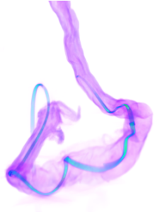
Credit: Jinxing Li, Zhenan Bao, Stanford University
Zhenan Bao, PhD, Xiaoke Chen, PhD, and colleagues from Stanford University demonstrate that NeuroString is a soft and stretchable graphene-based biosensing neural interface to monitor the dynamics of monoamine neurotransmitters, including DA and 5-HT, in both the brain and the gut of living animals. With tissue-like mechanical properties, the NeuroString can interface acutely with the GI mucosa and allows chronically stable and multiplexed neurochemical sensing in the mouse CNS.
While NeuroString has less sensitivity and selectivity than the latest genetically encoded fluorescence probes, such electrochemical methods are advantageous for translational use in humans. The authors state that further development will be dedicated to improving the sensor’s spatial resolution, selectivity, and multiplexity, integrating it with wireless electronics, and validating its long-term implantation performance.


