
April 15, 2018 (Vol. 38, No. 8)
Caroline Seydel Contributor GEN
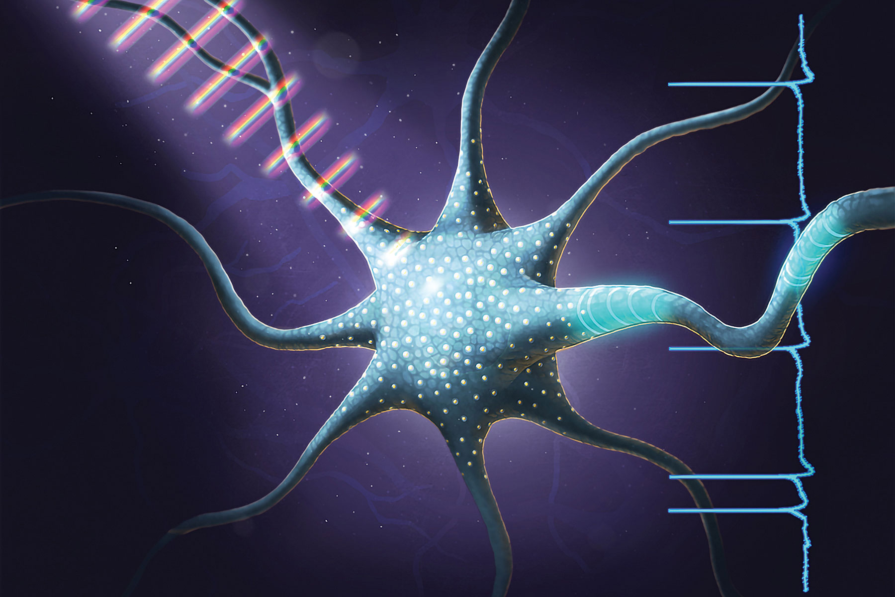
Optogenetically Triggered Processes Needn’t Become Visualization and Mapping Blurs
An optogenetic effect, like domino-toppling extravaganza, requires careful preparation. It also presents a chain-reaction sequence of events that may end all too soon. At least with dominoes, the individual tiles are easily seen, as are the patterns that are traced when the dominoes fall. Optogenetic patterns, however, are more obscure.
They consist of twitchy neurons that may spark this way and that as they send electrical signals through thickets of axons and dendrites.
To keep up with optogenetic events, observers rely on visualization and mapping tools. These optogenetic event–following tools, together with optogenetic event–preparation tools known as opsins, are advancing optogenetic neuroscience.
By equipping cells with opsins, or light-activated proteins, researchers can acquire optogenetic control, the ability to activate cells by using pulses of light. By using fluorescent dyes and high-resolution microscopy, researchers can visualize optogenetically stimulated cells. And by using mapping tools, researchers can discover which neurons talk to each other and how information travels around the brain.
These three areas—optogenetic control, visualization, and mapping—were covered at SPIE Photonics West 2018, a wide-ranging photonics, laser, and biomedical optics event in San Francisco that allotted a fair number of conference slots to neurogenetics, optogenetics, and optical manipulation. Several presentations highlighted optogenetic tools—molecules, microscopes, and software—that can improve our understanding of the interactions between living neurons in their native state.
Targeting Single Neurons
“If you look at two neurons that are next to each other, they’re often doing completely different things,” said Edward S. Boyden, Ph.D., professor of biological engineering and brain and cognitive sciences at the MIT Media Lab. While traditional optogenetics techniques activate or suppress whole populations of neurons in unison, that doesn’t mimic the living brain. Dr. Boyden sought a way to activate single cells with optogenetic methods. Because the long trailing processes of neurons overlap and intertwine, it’s difficult to stimulate just one cell of interest without also “zapping” its neighbors.
Dr. Boyden and colleagues solved this problem by fusing a powerful opsin, CoChR, to a small peptide that localizes the molecule to the cell body, so it’s not expressed in axons or dendrites. They teamed up with Valentina Emiliani, Ph.D., director of the Neurophotonics Laboratory at Paris Descartes University. Dr. Emiliani’s laboratory specializes in projecting three-dimensional holographic light into the brain. By designing a hologram that targets only the cells of interest, the Boyden/Emiliani team was able to activate just the neural networks they wanted to observe while avoiding crosstalk.
This ability to stimulate individual neurons and see which other neurons they activate opens up a world of possibilities. “You can target one at a time and map out the wiring,” Dr. Boyden explained. “If you could really understand the detailed patterns of brain activity and how they boost health or cause disease, we might be able to make ultraprecise new treatments.”
Another way Dr. Boyden’s laboratory zooms in on individual cells is by blowing them up. Called “expansion microscopy,” the technique is just what it sounds like: a biological sample is embedded in a polymer, which swells to many hundreds of times its original size in water. The expanding polymers carry the biomolecules along with them, and that makes it possible to resolve individual molecules that are normally far too densely packed to see.
“The applications are skyrocketing,” declared Dr. Boyden. His laboratory recently optimized the technique for looking at human tissue, which could pave the way for easier processing of biopsies. Furthermore, the laboratory found that an expanded tissue specimen could be re-embeded and expanded again, achieving almost single-molecule precision. “With a linear number of steps, you can exponentially magnify something,” Dr. Boyden asserted. “Imagine if every microscope on earth could do single-molecule imaging.”
Mapping the Brain’s Connections
Besides molecular biology or chemistry, some investigators are turning to mathematics to resolve the brain’s complexity. Stephen Boppart, M.D., Ph.D., is the head of the Biophotonics Imaging Laboratory at the Beckman Institute for Advanced Science and Technology at University of Illinois, Urbana-Champaign, and his laboratory has developed a computer algorithm to tease out the patterns underlying the mountains of neural data being collected.
“This information is going to help us figure out which neurons are connected—how they respond to one another,” Dr. Boppart said. The algorithm compares each neuron against every other neuron in the circuit to create “correlation maps.” These maps will show how patterns of firing and resting change over time and paint a picture of the overall dynamics of the circuit.
More precise neural circuit maps will allow finer control for manipulating the nervous system. On that front, Dr. Boppart’s laboratory has been working with specially designed ultrafast light pulses that control not only whether the neuron fires, but how it fires. “We’re tickling the receptor, not just flooding it with light,” he explained. “When we change the spectral properties of the pulse, we can cause the neuron to have either greater current or lesser current through its channel.”
As optical engineers, Dr. Boppart and colleagues came at the problem from a different angle. They asked: Instead of changing the molecular biology, can we change the light stimulus? This question led Dr. Boppart to envision a setup where instead of diffusely illuminating all the cells, investigators could specifically target just those neurons in the circuit.
The algorithm and the light pulses together create a feedback system to refine the map of neural connections. “We send in light to different cells the way that we want; we collect the video data of the cell activity; and we analyze it with our algorithm,” summarized Dr. Boppart. “Ultimately, our analysis can tell us how we should stimulate those cells differently to get the function that we want.”
At the University of Illinois, Urbana-Champaign, researchers used ultrafast pulses of tailored light to make neurons fire in different patterns. These researchers, led by Stephen Boppart, M.D., Ph.D., declare that their work marks the first time coherent control of the wavelength and intensity of sequential light pulses succeeded in modulating a living cell’s optogenetically stimulated functions. [Stephen Boppart, M.D., Ph.D.]
Repairing the Retina
Beyond basic research, optogenetics techniques are being applied to disease-damaged retinas in hopes of regaining lost function. Scientists at NanoScope Technologies have developed a “multicharacteristics opsin,” or MCO, that responds to low-intensity, ambient light, rather than laser light. By infusing these molecules into retinal cells, NanoScope scientists have restored light sensitivity to the eyes of mice with macular degeneration.
Neurons in the retina called bipolar cells typically convey the signals from the photoreceptor cells to the retinal ganglion cells. “There are plenty of bipolar cells, as compared to ganglion cells, and they are very close to photoreceptors,” said Samarendra Mohanty, Ph.D., founder and chief scientific officer at NanoScope. “Most of the signal processing of the retina is not lost.” By delivering MCO to the bipolar cells, the NanoScope scientists imbued them with the ability to react to light.
The challenge, Dr. Mohanty noted, is delivering the opsin to only those areas that need it. As the disease progresses, photoreceptor cells die off gradually: some continue to function while others atrophy. The usual delivery method, a viral vector, installs the opsin indiscriminately all over the retina. “You don’t want that,” Dr. Mohanty insisted.
“Because you have photoreceptors intact in the most part of the retina, you don’t want to have a new layer of photoreceptor cells,” he continued. “That one will interfere and distort the image.”
NanoScope developed a laser delivery system, similar to electroporation, that delivers the opsin into cells in only selected regions of the retina. Also, NanoScope developed an all-optical method for detecting retinal cell stimulation, eliminating the need for labels or dyes. When the neuron is activated by light, it undergoes an electrical change, and the change in refractive index can be measured by optical interferometry.
“Our interferometry is sensitive to hundred-picometer resolution,” Dr. Mohanty explained. “We can measure very, very low deflections in the cell.” This detection system could be used to confirm the presence of functioning opsins in the retinal cells, but it could also perform a diagnostic service, detecting the progression of retinal degeneration as photoreceptor cells fizzle.
Speeding 3D Microscopy Scans
Elizabeth Hillman, Ph.D., professor of biomedical engineering at Columbia University, has developed a new microscopy technique called swept confocally aligned planar excitation microscopy (SCAPE). It was recently licensed to Leica Microsystems.
SCAPE allows researchers to image living things, like a Drosophila larva, in real time without immobilizing the sample. With this technique, researchers can accomplish otherwise impossible tasks, such as observing an animal’s neurons while the animal is moving and reacting to stimuli.
Like light-sheet microscopy, SCAPE sends a thin plane of light into the biological sample, but the light enters at an oblique angle, rather than horizontally. Then the light sheet is reflected off a mirror, and the camera stays focused on the mirror. The image is captured by moving the mirror rather than moving the sample or the camera.
SCAPE can be combined with optogenetics and other tissue manipulations during imaging because, unlike other systems, it does not require any movement of the imaging objective lens or the sample to create a 3D image. Also, because the microscope scans the entire volume, the sample doesn’t need to be restrained, as it normally would.
“If we move the mirror 10 times a second, we get 10 volumes a second,” Dr. Hillman noted, “which is faster than any point-scanning system.” SCAPE achieves up to 100 times the imaging speeds of confocal or two-photon microscopy, without phototoxicity, “and there’s not very much standing in our way of going even faster,” Dr. Hillman added.
Neuroscience provides a perfect opportunity for testing the capabilities of the technology, because of the wide assortment of fluorescent indicators already in use.
“It’s just been a fantastic playground to be able to apply the method to a lot of different samples related to neuroscience,” Dr. Hillman said. Her team sees lots of potential applications for SCAPE, from blood vessel development to the study of bone deformation in microgravity conditions. Her team has even worked with a volcanologist to study bubbles as they move through flowing liquids that have solids in them. “What’s fun,” Dr. Hillman related, “is that now we have the pleasure of figuring out what on earth we can measure, now that we can do this.”


