
Camille Mojica Rey Contributing Editor Clinical OMICs
Single-cell Sequencing Techniques are Providing Significant Advances Across a Broad Swath of Fields
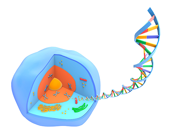
Until recently, scientists studying multicellular organisms at the cellular level had one big problem: their techniques did not allow them to account for cell-to-cell variation. That’s because the technology required the use of bulk tissue samples and results could only be interpreted as an average of the cells in a sample. But today, due to advances in analytic methods developed over the past 10 years, scientists routinely conduct research of individual cells.
“Single-cell analysis is transforming how scientists study biological systems,” said Alex Shalek, Ph.D., assistant professor of chemistry at MIT’s Institute for Medical Engineering & Science. “The cell is the fundamental unit of biology and, thanks to recent advances, we can now comprehensively profile a cell’s contents, its entire transcriptome,” said Shalek, whose lab focuses on developing innovative technologies. Single-cell analysis is empowering discovery by removing the need to a priori select the most important variables, thus eliminating bias. “I wouldn’t be surprised if single-cell approaches evolve to become the de facto standard for characterizing all biological specimens,” he added.
The work conducted today for basic research using single-cell analysis techniques will very likely be used to improve clinical diagnosis, treatment and monitoring. Shalek’s co-authored a paper in Science Translational Medicine last year stated that single-cell RNA sequencing (scRNA-seq), in particular, has the potential to “empower clinical implementation of personalized medicine.”
Currently, single-cell DNA sequencing (scDNA-seq) is not as easy to perform as scRNA-seq. That’s because a single cell can have hundreds of copies of a particular gene, while DNA only has two copies (one from each chromosome). Yet while scDNA-seq is in its infancy, scRNA-seq is in its adolescence, with multiple companies coming to market with technologies designed to create libraries for scRNA-seq (see Preparing scRNA-seq for the Clinic & the Field). Employing these, researchers can now prepare sequencing libraries of thousands or tens of thousands of cells. Leveraging scRNA-seq, researchers are addressing previously intractable problems, including characterizing the evolution of cancer and the attributes of tumor cells that cause therapeutic resistance, revealing the complexity of the nervous system, and honing the genes that allow a parasite to cause disease.
Source: Alfred Pasieka/Science Photo Library / Getty Images
Characterizing Cancer
Despite its promise, a lack of spatial-temporal context is one of the challenges to making the most of single-cell analysis techniques. For example, information on the location of cells is particularly important when looking at how a common form of early-stage breast cancer, called ductal carcinoma in situ (DCIS) progresses to a more invasive form, called invasive ductal carcinoma (IDC). “Exactly how DCIS invasion occurs genomically remains poorly understood,” said Nicholas Navin, Ph.D., associate professor of Genetics at the University of Texas MD Anderson Cancer Center. Navin is a pioneer in the field, developing one of the first methods for scDNA-seq.
Cellular spatial data is critical for knowing whether tumor cells are DCIS or IDC. So, Navin developed topographical single-cell sequencing (TSCS). Navin and a team of researchers published their findings in February 2018 in Cell. “What we found was that, within the ducts, mutations had already occurred and had generated multiple clones and those clones migrated into the invasive areas,” Navin said.
Navin and his colleagues are also using single-cell techniques to study how triple-negative breast cancer, becomes resistant to the standard from of treatment for the disease, neo-adjuvant chemotherapy. In that work, published in an April 2018 online issue of Cell, using scDNA-seq and scRNAseq, Navin and his colleagues found responses to chemotherapy were pre-existing, thus adaptively selected. However, the expression of resistant genes was acquired by subsequent reprogramming as a result of chemotherapy. “Our data raise the possibility of therapeutic strategies to overcome chemoresistance by targeting pathways identified in this study,” Navin said.
Source: SCIEPRO/Science Photo Library / Getty Images
Revealing Complexity
The authors of research published in 2017 in Genome Biology also identified lineage tracing as one of the technologies that will “likely have wide-ranging applications in mapping developmental and disease-progression trajectories.” In March researchers published an online study in Nature in which they combined single-cell analysis with a lineage tracing technique, called GESTALT (genome editing of synthetic target arrays for lineage tracing), to define cell type and location in the juvenile zebrafish brain.
The combined technique, called scGESTALT, uses CRISPR-Cas9 to perform the lineage tracing and single-cell RNA sequencing to extract the lineage records. Cas9-induced mutations accumulate in a CRISPR barcode incorporated into an animal’s genome. These mutations are passed onto daughter cells and their progenies over several generations and can be read via sequencing. This information has allowed researchers to build lineage trees. Using single-cell analysis, the team could then determine the diversity of cell types and their lineage relationships. Collectively, this work provided a snapshot of how cells and cell types diverge in lineages as the brain develops. “Single-cell analysis is providing us with a lot of information about small differences at cell type-specific levels, information that is missed when looking at the tissue-wide level,” said Bushra Raj, Ph.D., a postdoctoral fellow in Alex Schier’s lab at Harvard University and first author on the paper.
Raj’s collaborators included University of Washington’s Jay Shendure, Ph.D., and Harvard Medical School’s Allon Klein, Ph.D., pioneers in the field of single-cell analysis. The team sequenced 60,000 cells from the entire zebrafish brain across multiple animals. The researchers identified more than 100 cell types in the juvenile brain, including several neuronal types and subtypes in distinct regions, and dozens of marker genes. “What was unknown was the genetic markers for many of these cell types,” Raj explained. “This work is a stepping stone,” she added. “It’s easy to see how we might one day compare normal gene–expression maps of the brain and other organs to help characterize changes that occur in congenital disease or cancer.”
Raj credits single-cell analysis with accelerating the field of developmental biology.
“People have always wanted to work at the level of the cell, but the technology was lacking,” she said. “Now that we have all of these sequenced genomes, and now that we have these tools that allow us to compartmentalize individual cells, this seems like the best time to challenge ourselves as researchers to understand the nitty-gritty details we weren’t able to assay before.”
Building an Atlas
Human disease-relevant scRNA-seq is not just for vertebrates. For example, a team of researchers at the Wellcome Sanger Institute are working on developing a Malaria Cell Atlas. Their goal is to use single-cell technology to produce gene activity profiles of individual malaria parasites throughout their complex lifecycle. “The sequencing data we get allows us to understand how the parasites are using their genomes,” said Adam Reid, Ph.D., a senior staff scientist at the Sanger. In March 2018, the team published the first part of the atlas, detailing its results for the blood stage of the Plasmodium lifecycle in mammals. Reid contends these results will change the fight against malaria. “Malaria research is a well-funded and very active area of research. We’ve managed to get quite a bit of understanding of how the parasite works. What single-cell analysis is doing is allowing us to better understand the parasite in populations. We thought they were all doing the same thing. But, now we can see they are behaving differently.”
The ability to amplify very small amounts of RNA was the key innovation for malaria researchers. “When I started doing transcriptome analysis 10 years ago, we needed to use about 5 micrograms of RNA. Now, we can use 5 pico grams, 1 million times less,” Reid said. That innovation allows scientists like Reid to achieve unprecedented levels of resolution in their work. For Reid, increased resolution means there is hope that science will be able to reveal how malaria evades the immune system in humans and how the parasites develop resistance to drugs. Reid predicted the Atlas will serve as the underpinning for work by those developing malaria drugs and vaccines. “They will know where in the life cycle genes are used and where they are being expressed,” he said. Drug developers can then target those genes. The Atlas should be complete in the next two years, Reid added.
In the meantime, Reid and his colleagues are focused on moving their research from the lab to the field, particularly to Africa. “We want to look at these parasites in real people, in real settings, in real diseases states,” he explained. Having access to fresher samples is one reason to take the research into the field. “The closer we can get to the disease, the better chance we have of making an impact.” Reid anticipates that RNA-seq technology is on the verge of being portable enough to go into the field (see Preparing scRNA-seq for the Clinic & the Field). Everything from instrumentation to software is developing rapidly, he said. Reid also said that the methods used to understand the malaria parasite will likely be used to understand and create atlases for other disease vectors.
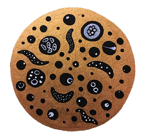
A gold leaf paint and ink depiction of the Plasmodium falciparum lifecycle by Alex Cagan.
Path Ahead
It is clear to those using single-cell analysis in basic research that the path ahead includes using the techniques in the clinic. “As the technologies become more stable, there will be a lot of opportunities for clinical applications,” Navin said. These include early detection by sampling for cancer markers in urine, prostate fluid, and the like. It also includes non-invasive monitoring of rare circulating tumor cells, as well as personalizing treatment decisions using specific markers. These methods will be particularly useful in the case of samples that today would be labeled QNS, or ‘quantity not sufficient.’ “Even with QNS samples, these methods allow you to get high-quality datasets to guide treatment decisions.”
This article was originally published in the May/June 2018 issue of Clinical OMICs. For more content like this and details on how to get a free subscription, go to www.clinicalomics.com.


