
September 15, 2018 (Vol. 38, No. 16)
Gail Dutton
Fluid Imaging Technologies Shows Biopharma How Flow Imaging Outshines Light Obscuration
This past spring, the U.S. Pharmacopeia indicated that it was considering new guidelines for the determination of particulate matter in therapeutic formulations. Existing guidelines emphasize the use of light obscuration technology, but they could be revised to reflect the growing importance of flow imaging technology, which offers advantages in analyzing biopharma formulations. Flow imaging, for example, excels at distinguishing protein aggregates from other contaminants.
Light obscuration has been the gold standard for the past 30 years, so a shift toward flow imaging would have to overcome considerable inertia. It happens that momentum favoring a shift to new technology has been building for at least two years, ever since the FDA requested that determinations of particulate matter exceed the customary 10-micron limit. Flow imaging provides resolution as fine as two microns.
A need for finer resolution has been recognized by Fluid Imaging Technologies, a manufacturer of particulate analysis instrumentation. “We’ll announce a new instrument combining light obscuration with flow imaging,” says Kent Peterson, the company’s president and CEO.
Finer resolution is possible for transparent and translucent particles, as well as for opaque. Fluid Imaging Technologies, which pioneered flow imaging, released the FlowCam Nano last spring to take detection to 300 nm with imaging and characterizing particles in a fluid sample. The increased resolution is particularly useful when particle morphology is critical for characterization. More than 40 physical parameters can be determined and analyzed from each image.
In one practical example, the FlowCam Nano allows the real-time identification of the 500-nm spirochete Borrelia burgdorferi, which causes Lyme disease. Previous detection methods required the more time-consuming serological testing, cultures, or PCR amplification. The FlowCam family of instruments is also used for process troubleshooting and quality testing.
Last February, Fluid Imaging Technologies won Pittcon Today’s Silver award for excellence for the FlowCam Nano and its Nano-Flow Imaging™ capability. The award recognizes new, ingenuous products for their implementation, outcomes, creativity, and expected impact in the field of laboratory science. “This validates the efficacy of the technology, as well as the innovative culture of the company and employees,” Peterson says.
Fluid Imaging Technology’s history of innovation began nearly 20 years ago, as a spinoff from Bigelow
Laboratory for Ocean Sciences (BLOS) in coastal Maine. It introduced a new concept in imaging—a digital imaging flow cytometer—from its headquarters above a garage. The device caught on quickly among oceanographic researchers, and the garage became a distant memory. As the company expanded to larger, more functional facilities, its applications also expanded, first into municipal water and later into the chemical, oil, and gas industries.
In 2010, following the research of John Carpenter, Ph.D., a professor of pharmaceutical sciences and co-director of the Center for Pharmaceutical Biotechnology at the University of Colorado Denver, who pioneered the use of flow imaging to study particles in biomaterials, Fluid Imaging Technologies entered the biotech market. Based on Dr. Carpenter’s papers, Peterson and his team realized that flow imaging had extensive applications in the pharmaceutical arena. The challenge, of course, was convincing everyone else.
The task, especially during the early days, was to educate people about the concept of digital imaging flow cytometry, Peterson recalls. “This was a new concept in imaging. The notion of combining static imaging, manual microscopy, and pure flow cytometry in one instrument, and performing the analysis in one fell swoop, was new to researchers.” The same challenges remain today, as researchers in new disciplines learn what flow imaging actually means in terms of the value it can bring to their labs.

Kent Peterson, president and CEO, Fluid Imaging Technologies
Imaging and Analysis Is an Advantage
“For pharma, flow cytometry isn’t the biggest need. Imaging and analysis is,” Peterson says. “There are many technologies to address all kinds of problems in developing pharmaceutical formulations. For example, six or eight address particle analysis—mainly focusing on determining particle sizes of calculating their concentrations in fluids. Ours is the only approach that provides images, which enable those particles to be identified,” Peterson says.
For formulation scientists, the technology enables early identification of the origins of colloidal activity and protein aggregation for injectable therapeutics and lyophilized biologics. With this information, they can better meet their goals for safety, efficacy, and shelf life.
For example, Peterson says, “knowing the number of particles is a start, but let’s say 1000 particles are present in a fluid. Maybe 300 of them are air bubbles. Of the remainder, what if half are silicone droplets, others are protein agglomerates, and a few are glass shards. Where did the silicone come from? The glass shards?” he asks. Flow imaging provides the resolution and the analysis that lets developers ask these questions. Studies show that many of these particulates are undetectable using light obscuration technology. The benefits of flow imaging, Peterson says, are “orders of magnitude improvements in the value proposition.”
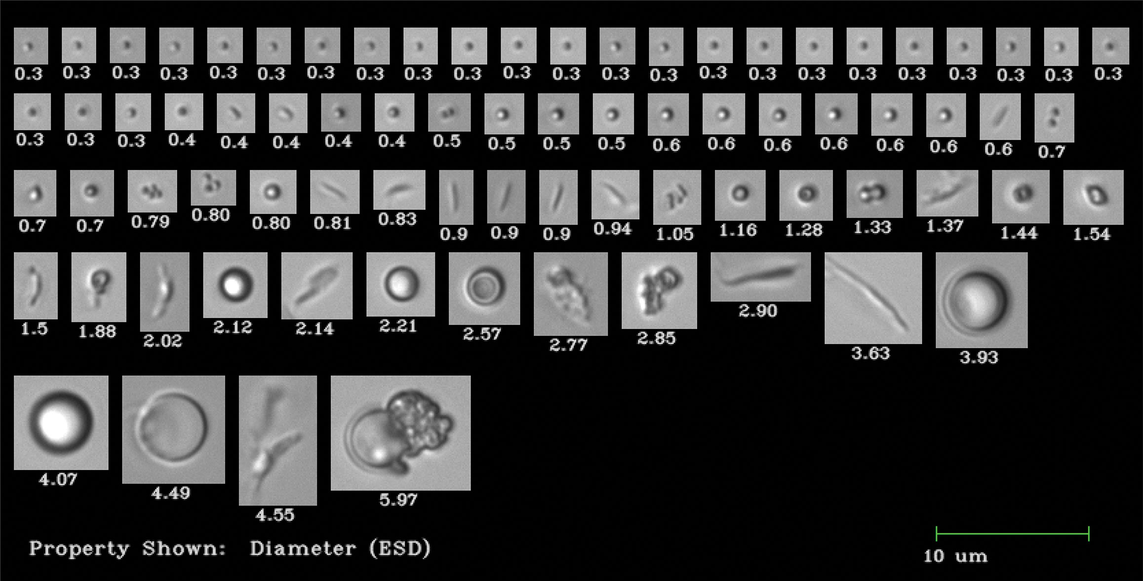
Fluid Imaging Technologies’ FlowCam system provides image-based analyses of the particles that occur in protein therapeutics. It allows biopharmas to simultaneously determine particle shape, type, and size distribution. The system also displays an image of each particle measured.
More Applications to Come
“We’re just scratching the surface of what this technology can do in biotech,” he continues. For example, Fluid Imaging Technologies is collaborating with a researcher to test and identify neonatal blood bacteria instantaneously, allowing the best antibiotic to be determined and administered within minutes. The time-consuming alternative to this technology is to culture the bacteria before determining which antibiotic to administer. “The researcher performing those studies has applied to the NIH for funding to advance the project,” Peterson says.
Another application aims to identify circulating tumor cells. “We’re in collaboration with a large, well-known institution to prove that capability. This could be a rapid, cost-effective way to monitor cancer treatment.” Improving the effectiveness of separating cells from magnetic beads is another potential application, Peterson suggests. He foresees many untapped areas with substantial upside for the company.
Fluid Imaging Technologies


