
Cell line validation has been under a bright spotlight since evidence emerged of widespread misidentification and contamination of cell lines, including those used in preclinical research and cited in thousands of papers. And preclinical research is not the only arena where misidentified or contaminated cell lines can wreak havoc and undermine years of work.
Failing to correctly validate cell lines used to produce therapeutic proteins can have substantial financial repercussions for biopharmaceutical manufacturers and, in extreme scenarios, pose safety risks for patients. In the manufacturing game, validation means verifying not only identity but also purity, sterility, and functionality in ways that meet regulatory agency standards.
Confirming Identity
|
Click Image To Enlarge +
|
 |
|
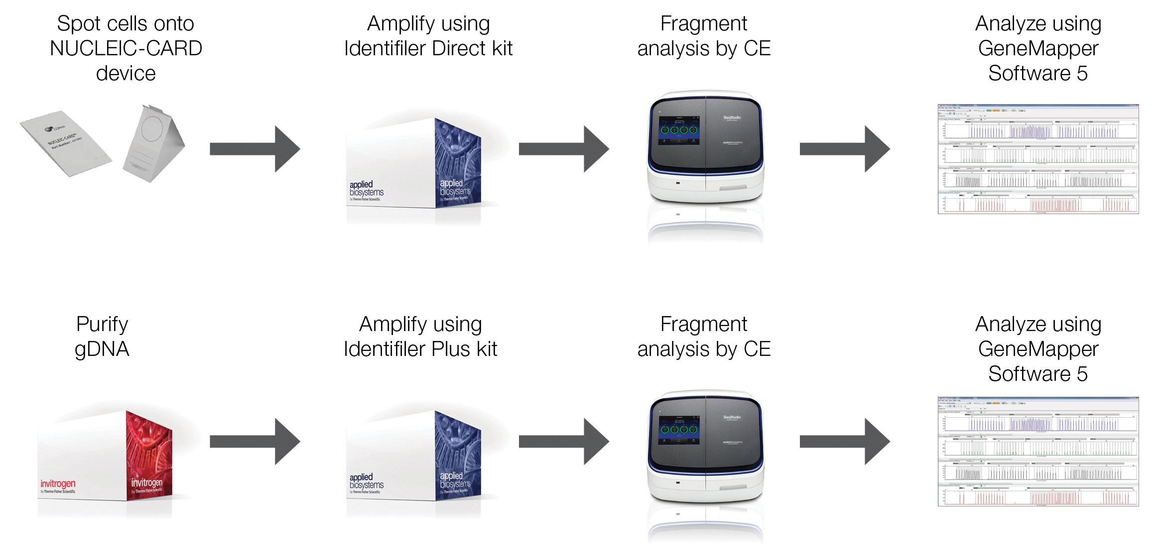
ThermoFisher’s GeneMapper® 5 Software allows users to process and compare short tandem repeat (STR) profiles between samples after co-amplification and fragment analysis of STR loci using an STR analysis kit and capillary electrophoresis, respectively. Having the capability to monitor changes in STR profiles over time can help researchers spot both contamination and genetic drift in their cell cultures.
|
Confirming cell identity doesn’t actually begin at the lab bench. “Documentation of the history of the cell line is just as important as the testing of that cell line,” explains Michael J. Hantman, Ph.D., associate director, methods development and validation, at Charles River Laboratories. “If you can’t say where the cell line came from—that’s going to be a big red flag for regulatory agencies.”
Once the manufacturer has collected adequate documentation on the history and characteristics of the cell line, it can apply a variety of techniques to authenticate cell identity—ranging from simple morphology checks, to classic karyotyping analyses, to modern gene sequencing methods.
Phenotypic methods, such as cell morphology and growth curve analysis, are perhaps the simplest methods used to validate cell lines. The American Type Culture Collection (ATCC) recommends frequent, brief observations of cell morphology, which can change depending on culture density, differentiation state, cell health, and media composition. Unusual morphology can indicate underlying issues—including contamination with another cell line or microorganism. Similarly, sudden changes in growth can also indicate a problem; it’s important to monitor cell growth for consistency using a growth curve analysis.
Isoenzyme analysis provides another relatively rapid and inexpensive method to identify species of origin and detect cross-contamination. Different isoforms exist for a number of intracellular enzymes, and isoenzyme analysis uses electrophoretic banding to reveal subtle differences in the structure and motility of these variants—creating distribution patterns unique to different species. One limitation is that isoenzyme analysis cannot distinguish between cell lines from the same species.
Regarding karyotyping, according to Dr. Hantman, it’s “a classic method but really informative.” Scientists can use the number and appearance of chromosomes to confirm species of origin and identify human cell lines with unusual chromosome patterns sometimes observed in lines researchers have used for a long time.
Turning to gene sequencing, advances in the technology have made this contemporary technique more accessible and affordable—leading to its adoption in a wide array of applications, including cell line characterization. The extent of sequencing can range from a single gene to the whole genome.
For species-of-origin analysis, DNA barcoding is a popular technique. Referred to as the “Barcode of Life,” the highly conserved region in the mitochondrial cytochrome oxidase subunit I (COI) gene provides a short sequence that allows differentiation between animal species, and an extensive database exists for species identification. However, similar to isoenzyme analysis, DNA barcoding cannot distinguish cell lines at the subspecies level.
At the opposite end of the spectrum, some companies have begun to use next-generation sequencing (NGS) to perform whole-genome or whole-exome sequencing on cell lines. The method is increasing in popularity as sequencing costs decrease because it provides a massive amount of data on the cell line. In contrast to DNA barcoding, whole-genome sequencing can differentiate between cell lines originating from the same species and closely related cell lines, but it requires a reference to perform a comparative analysis.
For identification at the subspecies level, “the industry is really hot for cell line authentication by short tandem repeat (STR) analysis,” says Dr. Hantman. Originally developed by forensics labs to aid in criminal investigations, STR analysis uses short, repeated DNA sequences for identification. The number of repeated units often varies between individuals, and each location on a chromosome where these repeats occur represents a different contour in an individual’s genetic fingerprint.
STR analysis has become the gold standard for authenticating human cell lines, and the availability of do-it-yourself kits from companies such as Thermo Fisher Scientific has increased accessibility. Thermo Fisher Scientific’s AuthentiFiler™, Identifiler™Plus, Identifiler™ Direct, and GlobalFiler™ PCRamplification kits contain primers for 9, 15, 15, and 21 different autosomal STR markers, respectively—as well as the sex marker, amelogenin. These, and similar kits, use fluorescently labeled primers optimized to allow co-amplification of multiple STR loci in a single PCR reaction.
Following amplification, researchers can perform size- and color-based separation and detection of STR loci using capillary electrophoresis and compare the results to reference profiles in databases to verify cell-line identity. Scientists can also create internal references using GeneMapper® ID-X and GeneMapper 5 software (Thermo Fisher Scientific).
The software also allows scientists to monitor longitudinal changes in STR profiles, which in combination with a broad range of STR markers, can detect genetic drift. With every division, cells can pick up genetic mutations, and these mutations can cause discrepancies between STR profiles. While regulatory agencies realize that genetic drift can cause an imperfect match, these discrepancies can cause uncertainty and complicate the interpretation of results.
A more substantial barrier to widespread use of STR analysis, however, is the current lack of kits, guidelines, and databases for nonhuman cells. While human cell lines have become popular among manufacturers, Chinese hamster ovary (CHO) and mouse cell lines are also commonly used in protein production. Unfortunately for biopharma, the FBI didn’t have much incentive for chasing down Chinese hamsters during the development of DNA fingerprinting.
The National Institute of Standards and Technology (NIST) has teamed up with the ATCC to form a consortium of 13 laboratories to expand the use of STR analysis to mouse cell lines. The Mouse Cell Line Authentication Consortium (MCLAC) selected murine cell lines because of their widespread use in both biomanufacturing and preclinical research. MCLAC members have tested 50 of the most commonly used mouse cell lines using a multiplexed PCR assay developed by NIST. The results of this work should be published later this year, says Jamie Almeida, bioassay methods group, NIST. “The consortium’s data will lead to validated STR profiles for the mouse cell lines tested,” she adds. “These profiles will be available on the NCBI BioSample database.”
Testing for Contamination
In cell line validation, identity must be confirmed as well as purity and sterility. Cross-contamination can compromise product yield, quality, and safety. One of the reasons it’s important to identify the cells used to manufacture therapeutic proteins has to do with a practice that is meant to protect against cross-contamination. Manufacturers select panels for adventitious virus testing based on the cell line species. Human lines will undergo testing for HIV and other viruses that infect human cells, whereas murine lines undergo a different battery of tests.
“If you don’t have clear identification of what the cell line is, you could potentially apply the wrong testing,” says Henry Chiou, Ph.D., associate director, cell biology, Life Science Solutions, Thermo Fisher Scientific. This could lead to a significant safety risk for patients, although Dr. Chiou admits that it’s an “extreme scenario.”
Adventitious viruses are not the only invisible contaminants that scientists working with cell cultures need to be wary of—mycoplasma bacteria, which even high-magnification, bright-field, or phase microscopy cannot detect, have become common contaminants. Mycoplasma often outcompete cells for essential nutrients without altering the turbidity or pH of the culture. Not only can contamination cause decreased cell growth and protein production, but some species of mycoplasma are pathogenic.
“GMP sites can be closed down for multiple months to clean out bioreactors and all surfaces purely because of mycoplasma detection. It’s a huge deal from a business perspective as well as a quality perspective,” says Jeff Hou, Ph.D., manager of cell culture development, BioProcess Sciences, Pharma Services Group, Patheon (part of Thermo Fisher Scientific).
Multiple methods exist for detecting mycoplasma contamination, including direct culture, indirect culture, and PCR assays. Considered the gold standard for mycoplasma detection, the direct-culture method uses a nutritionally enriched broth and carefully controlled environmental conditions to cultivate mycoplasma on agar plates. Both the characteristic “fried egg” appearance of colonies on the agar and a distinct color change in the broth indicate mycoplasma contamination.
The direct-culture method requires lengthy incubation periods, however, and it takes a minimum of 28 days to complete. In addition, while sensitive, it cannot detect mycoplasma species or strains that are notoriously difficult to grow in culture. Instead, scientists use the indirect culture test to detect noncultivable mycoplasma using fluorescent DNA stains, which bind to mycoplasma DNA and reveal contamination through extranuclear fluorescence.
PCR-based assays have become a popular technique for detecting both cultivable and noncultivable mycoplasma, and organizations such as the ATCC offer PCR-based mycoplasma detection services and kits. The ATCC’s Universal Mycoplasma Detection Kit can detect over 60 species of the tiny bacteria. “There are a couple of testing laboratories that have demonstrated the comparability between qPCR tests and the gold standard,” notes Dr. Hou.
Functionality Testing
Although cell line identity, purity, and sterility must be validated during the manufacture of protein-based therapeutics, the endgame is all about the product, and even whole-genome sequencing can’t guarantee the cell substrate will yield large quantities of functional, high-quality protein. Thus, the final step in validation assesses the cell line’s ability to consistently produce high-quality protein. “After you produce the protein, you need to test the mRNA level by qPCR, and you need to check the protein by Western blot using a specific antibody to make sure the protein is what you want,” explains Leon Song, M.D., director, GenScript. The next step, he says, is to check protein activity using a bioassay designed to test protein function.
Biopharmaceutical manufacturers play a different game than preclinical researchers—with different rules and referees. Validating cell line identity, purity, sterility, and functionality using methods that meet regulatory requirements ensures product success and patient safety. “No one assay will provide all of the answers that a manufacturer needs—a multimodal approach provides greater confidence that a particular cell line is authentic and will provide the functionality needed for efficiency, productivity, and safety,” says Dr. Hantman.


