
Researchers from UT Southwestern (UTSW) and collaborators report they have captured images of an autoantibody bound to a nerve cell surface receptor for the first time using cryo-electron microscopy. Their findings reveal the physical mechanism behind a neurological autoimmune disease and may lead to new treatments for autoimmune conditions.
The new study is published in the journal Cell in a paper titled, “Structural mechanisms of GABAA receptor autoimmune encephalitis.”
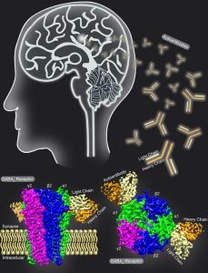
“Autoantibodies targeting neuronal membrane proteins can cause encephalitis, seizures, and severe behavioral abnormalities,” wrote the researchers. “While antibodies for several neuronal targets have been identified, structural details on how they regulate function are unknown. Here we determined cryo-electron microscopy structures of antibodies derived from an encephalitis patient bound to the γ-aminobutyric acid type A (GABAA) receptor.”
“We’re entering a new era of understanding how autoimmune disease works in the central nervous system,” explained Colleen M. Noviello, PhD, assistant professor of neuroscience at UTSW who specializes in obtaining cryo-electron microscopy (cryo-EM) images down to an atomic level of resolution. Noviello co-led the study with Ryan Hibbs, PhD, associate professor of neuroscience and biophysics, an Effie Marie Cain scholar in medical research, and an investigator in the Peter O’Donnell Jr. Brain Institute and Harald Prüss of Universitätsmedizin Berlin.
The team is planning to collaborate with colleagues to study more autoimmune conditions that affect the central nervous system.
“We identify key residues in these antibodies involved in specificity and affinity and confirm structure-based hypotheses for functional effects using electrophysiology,” concluded the researchers. “Together these studies define mechanisms of direct functional antagonism of neurotransmission underlying autoimmune encephalitis in a human patient.”

