Traditional reporter gene assays are powerful tools to interrogate adaptive stress-response signaling mechanisms. Such assays provide a convenient means for measuring global cellular responses to small molecule modulators. However, drug action typically occurs at signaling events far upstream of gene activation that are not directly assessed by genetic reporters. Furthermore, transcriptional readouts have limited utility for real-time pathway analysis, and often require extended drug treatment times for adequate performance. Consequently, alternate methods that measure signaling responses at upstream pathway nodes are desirable as complements to transcriptional reporters.
Stress-response pathways use a common pathway architecture involving dynamic regulation of protein turnover that allows for such alternate methods to be considered. For example, hypoxia-inducible factor-1A (HIF1A) pathway is acutely regulated by the ubiquitin-proteasome system. Under normoxic conditions, the transcription factor HIF1A is repressed by prolyl hydroxylation and VHL-directed ubiquitination. Upon transition to hypoxic conditions or chemical inhibition of HIF1A prolyl hydroxylation, HIF1A proteins accumulate resulting in HIF1A transactivation. The oxidative stress-response pathway is similarly regulated by protein lifetime.
In unperturbed cells, nuclear factor (erythroid-derived 2)-like 2 (NRF2) is subject to Keap1-directed ubiquitination and proteasomal degradation. Reactive oxygen species disrupt the interaction between Keap1 and NRF2, resulting in NRF2 accumulation and transactivation. While HIF1A and NRF2 mechanisms are key examples of the relevance of protein lifetime in regulation of gene activation, this pathway architecture is conserved within many signaling pathways. Thus, protein lifetime has the potential to be broadly exploited as a readout for critical upstream signaling nodes in adaptive stress response pathways.
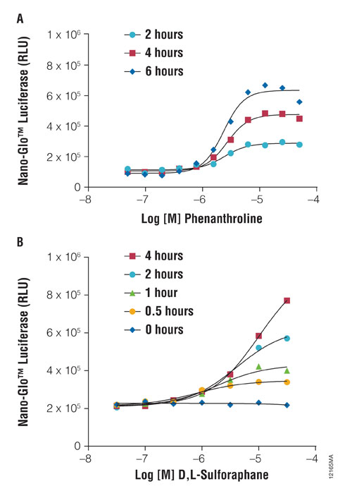
Figure 1. As a protein function reporter, NanoLuc Luciferase enables quantitation of changes in protein stability following induction of stress-response signaling. HCT-116 cells were transiently transfected with the pNLF1-HIF1A[CMV/neo] Vector and Transfection Carrier DNA and treated with hypoxia mimetic (A) or with the pNLF1-NRF2[CMV/neo] Vector and pKEAP1 DNA and exposed to a stimulant (B). Treatments occurred as indicated, and NanoLuc Luciferase was detected using Nano-Glo Luciferase Assay and a GloMax®-Multi+ Plate Reader.
Oxidative Stress and Hypoxic Responses
NanoLuc® luciferase is ideally suited as a protein function reporter for quantifying protein lifetime dynamics due to its small size (19 kDa) and intense bioluminescence output. Changes in intracellular protein levels can be quantified using Nano-Glo® reagent in a simplified assay format, wherein the luminescence output of NanoLuc luciferase is used as a surrogate for the intracellular levels of the fusion protein. It is possible to measure hypoxia or oxidative stress-response signaling via direct genetic fusions of NanoLuc luciferase with HIF1A or NRF2 proteins, respectively, in HCT116 colorectal cancer cells.
As shown in Figure 1, when HCT116 cells were transiently transfected with HIF1A-NanoLuc plasmid DNA, a chemical hypoxia mimetic (1,10-phenanthroline) induced a dose- and time-dependent accumulation of the reporter fusion. Similarly, an NRF2-NanoLuc fusion protein accumulated in HCT116 cells upon treatment with d,l-sulforaphane (a known inducer of reactive oxygen species).
Although protein stabilization is an initiating event in stress-pathway activation, a key outcome is the subsequent induction of genes required for maintaining cellular homeostasis. Multiplexed analysis of a NanoLuc protein fusion reporter with a traditional firefly luciferase transcriptional reporter allows for both signaling events to be analyzed in a single sample.
As an example, HEK293 cells were co-transfected with HIF1A-NanoLuc and hypoxia response element (HRE-luc2P) reporters, and the Nano-Glo Dual-Luciferase® Reporter Assay was used to sequentially assess both protein stability and transcriptional responses from the same cell population.
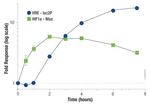
Figure 2. Multiplexed assessment of protein stability and gene activation in the same sample with the Nano-Glo Dual-Luciferase Reporter Assay. HEK293 cells were transiently transfected with the pNLF1-HIF1A[CMV/neo] fusion construct, diluted 1:1,000 into the Hypoxia Response Element Vector, pGL4.42[luc2P/HRE/Hygro]. After 18 hours, cells were stimulated with varying doses of 1,10-phenanthroline. At various time points, plates were assayed for expression of the firefly luciferase transcriptional reporter and the Hif1a-NanoLuc fusion protein using the Nano-Glo Dual-Luciferase Reporter Assay. The data shown represent the fold response for the 100 µM dose of phenanthroline relative to carrier alone; n = 4. Luminescence was measured on a GloMax-Multi+ Plate Reader.
As shown in Figure 2, HIF1A-NanoLuc protein accumulation peaked at approximately two hours post-stimulation, whereas eight hours were required for maximal induction of the hypoxia response element using the firefly luciferase reporter (luc2P). This application of dual-reporter assays allows for protein stability reporters to be readily multiplexed with transcriptional endpoints, enabling highly precise mode-of-action studies for toxicological screens.
Promega provides a suite of NanoLuc fusion vectors to enable intracellular protein stability assays. These vectors facilitate subcloning of N-terminal and C-terminal fusions with NanoLuc luciferase using standard restriction enzyme cloning or Promega’s Flexi® cloning system (Table). Flexi-system-compatible vectors facilitate transfer of the gene of interest from ORF clone gene panels, as provided within the Promega “Find My Gene” offering.
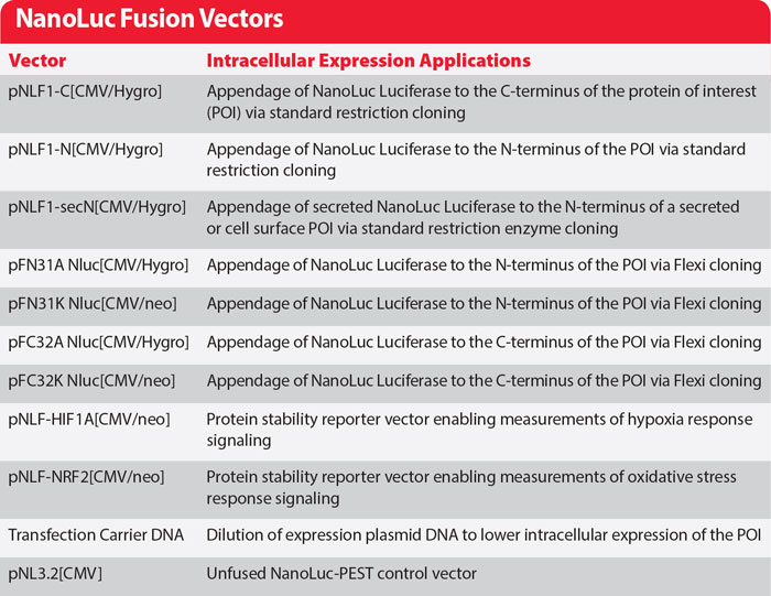
Promega’s NanoLuc vectors facilitate subcloning of N- and C-Terminal fusions using standard restriction enzyme cloning or the company’s Flexi Cloning system.
Matthew Robers ([email protected]) is a senior research scientist, Christopher T. Eggers, Ph.D., is a senior research scientist, Brock Binkowski, Ph.D., is a senior research scientist, Jim Hartnett is a senior research scientist, Jennifer Wilkinson is a research scientist, Chad Zimprich is a research scientist, Pete Stecha is a senior research scientist, and Mei Cong, Ph.D., is director of custom assay services at Promega.


