
January 1, 2006 (Vol. 26, No. 1)
Applying SureFire to Detect ERK 1/2 Phosphorylation
G protein-coupled receptors (GPCRs) represent a diverse group of signaling proteins and are involved in a wide variety of physiological processes. Of particular importance to the pharmaceutical industry, GPCRs, for the most part, have proved amenable as drugggable targets. As a result, GPCRs provide the largest group of therapeutic targets, and molecules that target GPCRs constitute a high proportion of pharmaceuticals on the world market.
Current methods employed in GPCR screening programs measure various cellular signaling events. The most widely used include measuring the formation of cyclic adenosine monophosphate (cAMP), inositol triphosphate (IP3), and intracellular Ca2+ mobilization. As a general rule, cAMP measurement provides useful data when screening receptors known to couple to Gi/o or Gs, whereas Ca2+ mobilization or IP3 formation assays are most useful for measuring activation of Gq/11-coupled receptors
More recently, the potential for using the phosphorylation of p42/44 extracellular signal-regulated kinase (ERK 1/2) as a measure of GPCR activation has become clear (Figure 1). A growing volume of evidence has shown that the signaling pathways activated by the stimulation of a wide variety of GPCRs converge at ERK 1/2 despite coupling to different G protein subclasses. In a screening context, this convergence at ERK 1/2 offers a broad spectrum GPCR functional screen.
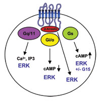
Fig 1: The major signaling pathways used by GPCRs, demonstrating the potential for ERK 1/2 phosphorylation with each major class.
SureFire Cellular Assay
TGR BioSciences (www.tgr-biosciences.com.au) recently developed the SureFire cellular ERK 1/2 assay. This assay is capable of detecting phosphorylation of ERK 1/2 in a cell-based format and allows the detection of not only Gq/11-coupled receptor activation but also many of the more difficult to detect Gi/o-coupled receptors and even some Gs-coupled receptors.
The naturally high intracellular concentration of ERK 1/2 means that no transfections, dye loading, or pre-stimulations are required to perform the assay. This, in turn, permits the generation of cell-based assay data that has a high degree of physiological relevance and allows measurement of ERK 1/2 activation in cells by endogenous receptors, including primary cells.
Because the assay is homogeneous and amenable to automation, the SureFire cellular ERK 1/2 assay can be used not only in lab-scale experiments but also in high-throughput screens performed by the pharmaceutical industry.
The SureFire cellular ERK 1/2 assay can be performed on cells grown in either 96-, 384-, or 1536-well microtitre plates. Generally, cultured cells are stimulated with the appropriate agonist for an optimal time, usually 515 minutes, and lysed with SureFire Lysis buffer.
For inhibition experiments, a similar protocol is followed, but the cells are pre-incubated with the inhibitor prior to stimulation. To determine the extent of ERK 1/2 phosphorylation, the lysed samples are simply activated’ with SureFire Activation buffer and incubated with SureFire Reaction buffer containing AlphaScreen beads (PerkinElmer) for two hours, at room temperature.
The signal in the wells can then be measured using a plate reader, capable of AlphaScreen measurements (Figure 2). There are no washing steps required and a minimum of liquid handling, affording easy automation and reduced handling errors.
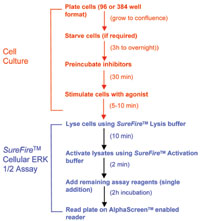
Fig.2: Outline of the standard SureFire cellular ERK 1/2 assay protocol. No washing steps are required, and from cell lysis to reading plates, takesless than 2.5 hours
Optimizing Phosphorylated ERK 1/2 Signal
Several factors can affect the level of ERK 1/2 phosphorylation within cells. In most cases, problems caused by low-signals, high-backgrounds, or signal variability can be remedied by simple optimization of cell culture procedures. Low-background levels of phosphorylated ERK 1/2 are crucial to achieving a good, uniform response in cells and high-signal background windows.
In our laboratories we have found several approaches to achieve low-backgrounds. Confluent monolayers of cells can promote contact inhibition with consequent reduction in background levels of ERK 1/2 phosphorylation. Removal of serum prior to assaying by cell starvation can lower background levels, but serum removal is not mandatory if the cells are grown for a few days and are confluent.
The formation of a confluent monolayer of cells also has the effect of reducing signal variability. Other factors that can affect assay performance include stimulation time, which can vary according to the receptor system but is usually maximal for ERK 1/2 phosphorylation between 515 minutes. The cell line used can also influence the efficiency of G protein-coupling to ERK 1/2; for example, a receptor system that gives a strong signal in CHO cells may not in HEK 293 cells.

Fig.3a: Dose response curves generated for all of the major GPCR G protein-coupling pathways, Gi/o(Muscarinic M4 receptor), Gq/11(PAR-1), and Gs(VAPC-1).
Assay Applications
Screening GPCRs via ERK 1/2 has several advantages. Most obviously, phosphorylation of ERK 1/2 provides a direct physiological functional readout of activation of Gi/o-coupled GPCRs. Currently, the most common method of achieving this is through screening for inhibition of adenylate cyclase activity by measuring the reduction of forskolin-stimulation of cAMP levels.
In contrast, many Gi/o-coupled receptors will activate ERK 1/2 directly through the Ras/Raf pathway, or via transactivation of the EGF receptor. Therefore, the SureFire cellular ERK 1/2 assay offers a simple and direct measure of Gi/o-coupled receptor activation.
In addition, activation of Gq/11-coupled GPCRs will also cause a consistent increase in the phosphorylation of ERK 1/2. Therefore, most or all receptors that can currently be screened using existing technologies, such as Ca2+ mobilization or formation of IP3, may also be screened using the SureFire cellular ERK 1/2 assay.
Gs-coupled receptors stimulate adenylate cyclase activity and are readily detected by assays that detect the formation of cAMP. However, certain Gs-coupled receptors can also give rise to increases in the level of phosphorylated ERK 1/2, although, the mechanism of the increase is not always clear.
Taken together, the SureFire cellular ERK 1/2 assay provides a powerful tool for the measurement of activation of many GPCR classes (Figure 3).
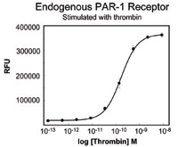
Fig. 3b:Cells were stimulated for between 5-10 min and subsequently lysed.
Orphan GPCR Screening
An added feature of the assay is that when combined with a cell line that offers low levels of endogenous ligand-induced ERK 1/2 phosphorylation, such as HEK 293 cells, the system can be useful for screening orphan receptors. HEK 293 cells show low levels of ERK 1/2 phosphorylation in response to many common agonists, including serum.
However, when receptors are transfected into HEK 293 cells, a strong agonist-mediated signal is observed (Figure 4). This offers a unique system for GPCR orphan receptor screening, because so few endogenous ligands activate ERK 1/2, and a broad array of transfected receptors can activate the ERK 1/2. Thus, no knowledge of the GPCR coupling mechanism is required.
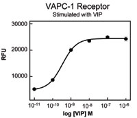
Fig. 3C: Cells were analyzed for phosphorylated ERK 1/2 using the SureFire cellular ERK 1/2 assay.
Assay Troubleshooting
While the measurement of ERK 1/2 phosphorylation offers a single screening platform for detection of GPCR activation, not all receptors behave as expected. We have tested many Gq/11-coupled receptors, all of which stimulate ERK 1/2 phosphorylation.
However, the success rate with Gi/o-coupled receptors is about 70% of receptors tested, and can be dependent on the cell line used for transfection. The mechanism of Gs-coupled receptor activation of ERK 1/2 is unclear, as is how widespread the phenomenon is.
The SureFire cellular ERK 1/2 assay detects ERK 1/2 phosphorylation in cells with a sensitivity equivalent to a Western blot. However, some receptors, such as those that couple to Gs, may have a low coupling efficiency to ERK 1/2. This may limit the use of ERK 1/2 as a screening tool for such receptors.
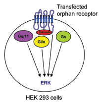
Fig. 4: Orphan receptor screening in HEK 293 cells using ERK 1/2 as an analyte.
Summary
The SureFire cellular ERK 1/2 assay offers a new approach to successfully screen many different receptor types and classes. Importantly, in addition to Gq/11-coupled receptors and certain Gs-coupled receptors, it provides an easy and physiologically relevant means of screening Gi/o-coupled receptors and adds to the tools available for GPCR research.


