
November 1, 2016 (Vol. 36, No. 19)
Richard A. A. Stein M.D., Ph.D.
Routes to Timely Diagnoses and More Effective Therapies for Neurological Disorders Are Emerging
Throughout the history of functional morphology, the brain was less forthcoming than many other organs. The brain was particularly challenging to early researchers, who often had to resort to the study of anomalies—illness- or trauma-related morphological changes, which might be found only in single patients, or in the cadavers of said patients.
Only in such unusual circumstances did the brain seem amenable to investigations that could relate structural features to distinct functions. For example, in the mid-1800s, based on autopsy observations in a patient who was able to utter only one syllable, Paul Broca described the involvement of the inferior frontal gyrus in speech.
“The brain is unique in its complexity,” says Gregory K. Farber, Ph.D., director, Office of Technology Development and Coordination, National Institute of Mental Health, National Institutes of Health. “We understand surprisingly little about basic operation in the central nervous system. Recent studies, however, have provided data of incredible quality.”
One such study is the Human Connectome Project, a multi-institutional research program that was launched in 2010 to expand knowledge about anatomical and functional connectivity in the brain. The Human Connectome Project was initiated as a five-year effort and drew on the work of two institutional consortia.
One consortium, led by Massachusetts General Hospital and the University of California at Los Angeles, used advanced magnetic resonance imaging to map fibrous connections in the brain. The other consortium, led by Washington University in St. Louis and the University of Minnesota, enrolled a population of 1,200 healthy young adults, including twins and non-twin siblings, in a study of the heritability of brain characteristics.
“The over arching focus of investigations as part of this project was on connectivity,” informs David C. Van Essen, Ph.D., a neuroscientist at Washington University. “The investigators involved realized that it would benefit the scientific community to have a broad attack on the questions of how the brain is wired and how this wiring relates to brain function and the control of behavior.”
Using a subgroup of participants in the Human Connectome Project, a recent study from Dr. Van Essen’s group addressed a conundrum that has for a long time intrigued biologists, the accurate mapping of the cerebral cortex. “What drew us into this effort,” recalls Dr. Van Essen, “was the need to understand how the brain in general and the cerebral cortex in particular is organized into distinct parcels or areas, which has been a century-old challenge in neurosciences.”
Like the cartographers of old, the scientists in the Human Connectome Project faced a daunting task. Although the cartographers were interested in geography, and the scientists were focused on the cerebral cortex, both were involved in mapping. The presence of multiple layers of organization is another commonality between the two.
“We have geographic maps that show the undulations of hills and valleys and describe the various features of the landscape,” explains Dr. Van Essen. “Overlayed on those maps are maps of political subdivisions, which include countries of varying sizes and shapes.”
To map the areas of the cerebral cortex, Dr. Van Essen and colleagues took advantage of a new methodology called multiband or multislice imaging. It provides more data per unit of time, higher quality data, and better resolution in space and time.
In the mapping study led by Dr. Van Essen, multiband imaging was used to identify 180 areas per cerebral hemisphere. Of these areas, 83 had been previously identified in postmortem studies, and 97 were completely new areas or subdivisions of known areas. “Investigators in the field have not reached a consensus as to how many areas there are in the cerebral cortex,” notes Matthew F. Glasser, Ph.D., the first author of the study and a medical student at Washington University.
“This is analogous to identifying the countries, as opposed to describing just the folding patterns, which would correspond to the mountain and valley terrain of the earth surface on geographical maps,” states Dr. Van Essen.
As part of this study, Dr. Glasser, Dr. Van Essen, and colleagues trained a machine-learning classifier to recognize the newly characterized cortical areas in new subjects, that is, subjects who did not participate in the original study. “[We showed that] investigators can relate their brain imaging results to this map, and that they can see how the results from different studies relate to each other,” asserts Dr. Glasser. The classifier was able to detect over 96% of the cortical areas in new subjects, and to correctly locate atypical topological arrangements that were present in some of the individuals examined.
“One of the most straightforward ways that this machine-learning algorithm could be useful is to help neurosurgeons who want to avoid injuries to specific brain areas that are important for certain tasks,” emphasizes Dr. Glasser.
The classifier and the parcellation map, which will be freely available to investigators, promise to provide a more sensitive tool to study the cerebral cortex and perform comparisons across individuals. “We anticipate that having better parcellation maps will help investigators explore processes during development and disease, and do so with more sensitivity and better localization,” concludes Dr. Van Essen. “This is the fundamental advance provided by our cortical parcellation.”
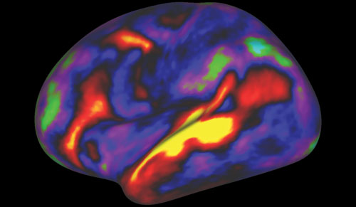
In this image of the brain’s left hemisphere, colors show activation patterns for people who were assigned the task of listening to stories. Active regions appear in red and yellow; inactive regions, blue and green. The image, from the Human Connectome Project, is a composite of MRI scans taken from a large population of healthy young adults. [Matthew F. Glasser, Ph.D., David C. Van Essen, Ph.D., Washington University]
Tying Up Loose Ends
“In the Human Connectome Project, the biggest concerns pertain to the uniformity of its data sources,” says Dr. Farber. “The data come only from healthy young adults, and we do not understand yet how the connectome changes over the lifespan.”
If successive studies were to recruit participants belonging to different age groups, from very young to elderly individuals, it would be possible to enhance our understanding of the connectome. Such studies are already being supported by several institutions including the National Institutes of Health.
“It will give us a huge amount of information to try to see how connectivity changes over time,” explains Dr. Farber. “It will be useful and necessary because many illnesses of the central nervous system start to occur from teenage years.” Several lifespan Human Connectome Projects are currently underway. “These studies,” predicts Dr. Van Essen, “will provide treasure troves of data and information for the field to share.”
Another challenge is that many of the data sources generated as part of the connectome studies have been established using noninvasive technologies. These technologies provide macroscopic views that may lack sufficient resolution. “Within every voxel of an MRI image, there can be millions of neurons,” notes Dr. Farber.
In many biological systems, mechanistic answers have been provided only by using cellular and molecular biology approaches, which historically have been more difficult to perform in the neurosciences, particularly for studies involving the cerebral cortex.
“Many of us think that true answers would come from an intermediate stage, between imaging and molecular techniques,” comments Dr. Farber. “Although the development of these intermediate-scale technologies is not there yet, this approach will almost certainly be needed to obtain a real understanding of what is going on.”

The connectome, a comprehensive map of all the neural connections in the brain, is represented by this abstract image of human brain. The image highlights just a few nodes and line segments to suggest how a connectome study may focus on selected portions of a complete wiring diagram. [Mirexon/Getty Images]
Links to Schizophrenia
“A lot of people tend to have their first episode of depression or schizophrenia during their late teenage years,” says Kirstie J. Whitaker, Ph.D., research associate in the department of psychiatry at the University of Cambridge. “We are not really sure why.”
Perturbations in several cortical areas might be involved in the pathogenesis of neuropsychiatric conditions, and interrogating the connectome emerges as a promising strategy to develop early diagnostic approaches and more effective therapeutic interventions.
“There are many different ways to build a connectome,” explains Dr. Whitaker. In two recent studies, Dr. Whitaker, Dr Vértes, Prof Bullmore, and colleagues from the Neuroscience in Psychiatry Network (NSPN) Consortium used two approaches to interrogate the connectome in human volunteers. “In both studies, we were able to gain insight into the biology of what is going on in specific brain regions, and we have accomplished that at a level that has not been done before.”
One study, led by Dr. Petra Vértes, an MRC fellow in bioinformatics at the University of Cambridge, compared functional MRI network parameters with maps of whole genome expression data available for the anatomically corresponding brain regions.
Using a reductionist approach to define gene expression profiles that appeared to be optimally predictive of the functional MRI parameters, the investigators defined two network phenotypes: one with high intra-modular connections and short distances between the connections, and one with low intermodular connections between cortical modules and long distances between connections.
The analysis revealed that integrative hubs, which mediate more long-distance connections, have higher metabolic requirements than hubs that mediate short-distance connections. This difference between the hubs indicates that integrative cortical areas are metabolically more expensive.
A second strategy used by Dr. Whitaker and colleagues, called structural covariance matrix, involves pairwise comparisons in the cortical thickness between cortical regions. Many studies revealed that cortical thickness typically decreases from teenage years to old age, with the fastest rates of shrinkage being observed during adolescence.
In a group of teenage and young adult volunteers, Dr. Whitaker and colleagues used MRI to measure cortical thickness and intracortical myelination. This analysis showed that in individuals who were around 14 years of age, the association areas were thicker and less myelinated than primary cortical areas, with the thinnest areas being found in somatosensory and visual cortices. Nonetheless, the rates of shrinkage increased during adolescence, and this was related to an increase in myelination.
Based on these findings, Dr. Vértes showed that gene transcription changes pointed toward the involvement of several genes known to modulate the risk of schizophrenia in this consolidation process of the association areas. These findings opened the possibility that during adolescence, deviating from the cortical consolidation trajectory in association areas, which are enriched for network hubs of the brain connectome, could explain the increased risk of schizophrenia historically found in this age group.
In the generation of these results, the availability and analysis of interdisciplinary and cross-disciplinary datasets have played an instrumental role. “Data has become available to allow investigators to improve the understanding of biology. And we need statisticians, computational modelers, and biophysicists to work with the biologists to generate data across all these different fields and start putting things together,” insists Dr. Whitaker.
Wiring Diagrams
“We are interested in addressing how a static wiring diagram can support the huge range of mental dynamics observed in humans or animals,” says Danielle S. Bassett, Ph.D., associate professor of bioengineering at the University of Pennsylvania.
Many previous analyses have pointed toward a static connectome, and human, mouse, and fruit fly studies have revealed that the wiring diagram of the connectome does not change very much during adulthood. However, it has been challenging to understand how a static wiring diagram can nevertheless support the functional fluidity of the brain and the transition between complex cognitive states.
One approach used by Dr. Bassett and colleagues to address this question takes advantage of applied mathematics. With this approach, Dr. Bassett’s team can analyze the organization of multiplex networks in the brain and examine how they support a variety of complex functions such as memory, language processing, and cognition.
“The second way in which we are trying to address this challenge is by a field called network control theory,” adds Dr. Bassett. This approach, which examines how a system whose components are organized in a complex interconnected network can be controlled, can provide mechanistic predictors of the dynamics of the system.
In a recent study, Dr. Bassett and colleagues used the framework provided by network control theory to predict, for specific wiring diagrams, the types of brain states and brain transitions that can be anticipated and the way in which transitions between different cognitive states could occur. The investigators also interrogated cortical areas that are most critical in controlling these transitions.
Although much research has been conducted on healthy adults, there is a gap in understanding wiring diagrams in disease states. Collaborative work between Dr. Bassett’s laboratory and other groups found evidence for multifocal dysconnectivity in specific cortical areas in young individuals with psychosis-spectrum disorders.
“We found that it is not one particular area of the brain that is affected,” reports Dr. Bassett. “Instead, whole circuits are altered.”
Hallmarks of this dysregulation included hyperconenctivity in certain areas and diminished connectivity in others, mirroring changes that have long been documented in adults with psychotic syndromes. “We need a lot of interdisciplinary cross-talk to better understand brain dynamics and to predict how to change the course of the system when something goes awry,” states Dr. Bassett.


