
January 15, 2009 (Vol. 29, No. 2)
Kathy Liszewski
As the Field Grows and Matures, It Is Becoming an Essential Tool in Product Development
Applications and products utilizing microfluidics are gaining momentum. The technology offers clear advantages in reducing reagent consumption, increasing speed and analytical performance, multiparameter testing, and user friendliness. The market is forecasted to grow to $1.9 billion by 2012, with the majority of revenue from diagnostic applications.
The “Lab Automation Conference” to be held in Palm Springs this month will showcase advances that include improvements in chip-to-world interfaces with standard geometries such as microscope slides and microwell plate formats, multiplexing capabilities, and off-the-shelf components for quick, low-cost implementation of microfluidic technologies.
Although lab-on-a-chip technology has been around for more than 20 years, its full promise has yet to be realized, says Holger Becker, Ph.D., founder and CSO of microfluidic ChipShop. “Many pioneering companies didn’t do well, and most laboratories do not yet incorporate microfluidics in their workflow,” Dr. Becker says. “We considered what we have learned from the microfluidics market in the last 10 years and how we could make this a more widely accepted and used technology.”
The company decided that more off-the-shelf options were needed. “For many years, if you wanted to incorporate microfluidics, it was necessary to go through an engineering project with the associated cost, time and development risk. Our idea was to provide an affordable start to microfluidics. We developed a two-pronged approach. First, we developed a tool box-like approach in which we offer tailor-made components. Second, we designed these to interface with existing lab equipment.”
Dr. Becker says the company now offers approximately 300 different chips with simple fluidic structures. “Companies can easily get hands-on experience for less than $300 and in a short time develop quick and dirty experiments to test their ideas. Then scientists can go to their boss to describe what works and what needs optimizing and how they can enhance success by building only a comparatively few structures.”
One of the big advantages of this component-driven approach is that one can use existing automation. “We built these components for classical applications such as electrophoresis,” Dr. Becker notes. “Also, users can use their existing robotics because the external geometry of the components is standardized into two common formats: a slide footprint or a microtiter plate format. This allows most users to engage their existing robotics platforms.”
For companies planning on integrating microfluidics into their research strategy, Dr. Becker has some advice. “It is best to speak to manufacturers as early in the process as possible. You need to be sure to develop the best design or it may be flawed and, ultimately, far too expensive. Your technology must be both functional and manufacturable at reasonable costs.”
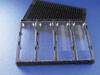
Modular microfluidic toolbox approach: Individual microfluidic chips (here depicted for continuous-flow PCR) the size of microscopy slides sit in a holder frame identical in geometry to a microtiter plate.
Multiplexed Clinical Analysis
Many microfluidic technologies are designed with the idea of one device for one application. This can be a problem, according to Vincent Gau, Ph.D., president of Genefluidics. “To assay something else, you would need another type of system. Our approach is different. We developed a universal microfluidic cartridge that can measure DNAs, RNAs, proteins, and small molecules.”
The key to the device is a two-component cartridge system in which the top portion stores reagents, waste, and sample, while the bottom portion houses microfluidic channels, valves, and vents as well as the integrated sensor array. The raw sample is loaded into the top portion and the assembled cartridge is inserted into the instrument that processes and analyzes the sample. Electrochemical detection methods allow ultra-high nontarget amplified sensitivity.
Dr. Gau notes that the gold standard for most types of DNA/RNA analyses is PCR amplification of a sample whose nucleic acid was first extracted and purified. “Our devices differ because we directly take a specimen such as blood and saliva and process that to completion within 10–20 minutes. The read-out has exceeding high sensitivity because we developed our own sensors that detect proportionally to the number of target molecules.”
Another important feature of this new approach is the ability to multiplex. “We are working with VA hospitals in Los Angeles and Palo Alto on urinary tract infection screening and the UCLA hospital on salivary diagnostics for oral cancers, comments Dr. Gau. “There are many cancer and disease biomarkers relating to RNA, DNA mutations, and proteins. Often, these are evaluated singly via PCR or an ELISA if it is a protein biomarker. However, with the universal cartridge system you can characterize any combination of desired markers, not just one biomarker. We anticipate making a custom blend and a universal panel that would be informative to the physician.”
The company will launch its devices in early 2009 for academic researchers and expects to work with strategic partners to develop other specific applications especially for multiplexing.
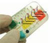
This handheld point-of-care system from Genefluidics has a universal sample-processing cartridge for conducting multiplexed assays and is field deployable.
UV Imaging Detection
Identifying and quantifying sample images in microfluidics technologies can be a challenge and a bottleneck. Paraytec has a solution—an ultraviolet imager for use with microfluidic devices.
“Traditional optical detection technology revolves around the use of microscopes, which suffer from field of view but are excellent at gathering light,” according to Jim Lenke, engineering manager. “Microscopes are frequently connected to digital cameras for collecting and digitizing data. However, these data images take time to be converted directly into absorbance units (AU) and suffer from not having an available reference for absolute AU values. Thus, researchers struggle to get quantitation in microfluidic devices. Additionally, because volumes are exceedingly small other techniques such as fluorescence are often used to collect even more light signal.”
Paraytec offers its ActiPix™ brand of ultraviolet (UV) area imaging detectors that use sophisticated optics and patented signal referencing with Active Pixel area sensors for high sensitivity UV detection in microfluidics and other applications.
How is this new technology used in microfluidics? “Our ActiPix D100 UV area imaging detectors use an Active Pixel Sensor based imaging chip,” Lenke says. “We can view a 9 mm wide by 7 mm high area, which covers a large area of real estate in a microfluidic device. This is unlike a typical microscope arrangement whereby a selected small region of the channel is chosen as a detection area. As the image is an area pixel array, we have the ability to simultaneously measure absorbance in several channels.”
“The imager is part of a family classed as Active Pixel arrays,” he adds. “Modern-day digital cameras expose us to pixel technology. Pixels are really just tiny photon detectors arranged in an array. These are different from the well-known CCD chips where selectively chosen pixel regions or columns are difficult to isolate. Some performances of pixels between the two classes are different as well. Our imager is 1,280 columns wide by 1,024 rows high and each pixel is 7 by 7 micrometers. Only rows needed for sensing are used while other rows are switched off during that operation. Reducing the total number of rows needed vastly increases readout rate and data size.”
An ActiPix multiplexed capillary cartridge from Paraytec
Miniature Cell Culture
Utilizing microfluidic devices for cell analysis is a new strategy holding great promise for many applications ranging from drug discovery to toxicity testing, according to Philip J. Lee, director R&D at CellASIC. “Microfluidics can be used to engineer microenvironments at the cellular size scale with miniaturization and automation for mimicking the physiological environment of cells and tissues. This capability allows exposing cells to changing flow environments to obtain a signal-response not previously possible.”
The concept is based on using disposable, application-specific microfluidic plates and a universal flow control interface. The microfluidic plates hold sample solutions, media, and cells formatted with a microtiter plate footprint. A key feature is a perfusion barrier to control nutrient transport and cell localization. A glass coverslide bottom and transparent optical path allow for use in an inverted microscope or standard plate reader.
“We are using this system to study liver toxicity as a predictive model of drug effects,” says Dr. Lee. “The current limitation with using cultured cells in toxicity studies is getting them to behave similarly to cells in vivo. We are, however, overcoming these hurdles using microfluidic cell cultures that allow maintenance of primary liver cells for use in high-throughput automated analysis.”
The novelty of the automation-compatible system is that it “provides a design environment that mimics in vivo processes such as higher cell densities, cellular transport, and continuous perfusion. We built this system to fit average users in a typical biological workplace. Also, it is compatible with existing equipment and assays. Overall, this system allows a higher quality data at reduced costs.”
Because hepatocytes are the best understood tissue model, the company’s initial applications for the microfluidic cell culture system will use these cells for both dose-response toxicity testing and for drug-metabolism studies.
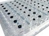
An automation-compatible 96-well array specifically designed to promote liver-specific activity of primary hepatocytes.
Enzymatic Assays
Much of drug discovery focuses on finding inhibitors of enzyme targets such as kinases, phosphatases, and proteases. Caliper Life Sciences offers its Mobility Shift Assay to interrogate druggable enzymes. It combines the advantages of capillary electrophoresis with microfluidics technology for the direct measurement of substrate and product.
Raj Singh, Ph.D., director of assay R&D, reports that the basis of the technology is Caliper’s sipper chip. “Our mobility shift microfluidics assay technology monitors the modulation of enzyme activity by inhibitors using a ratiometric fluorescence readout of the substrate and product. Basically, potent drugs produce a lower amount of the enzymatic product at a lower concentration than less potent ones. For example, kinase assays are not only analyzed on our LabChip EZ Reader, they also are rapidly profiled for selectivity and specificity against a panel of 200 plus enzymes.
“The assay components and compounds assembled in a microtiter well format are sipped into a 4- or 12-sipper chip via capillaries under vacuum. Reactions are separated by electrophoresis and signals measured by fluorescence in either endpoint or kinetic modes. The Mobility Shift Assay technology is designed for in vitro enzyme assays used in the drug discovery process. Even GPCR assays have been recently adapted to the Labchip format by utilizing the b-galactosidase enzyme fragment complementation technology commercialized by DiscoveRx as Pathhunter™.”
Dr. Singh says one of the big advantages of the technology is that it allows multiple read-outs from the same microtiter well without using any ancillary reagent or terminating the reaction. “Other technologies often use different methods for different drug discovery targets. Because the microfluidics technology only needs nanoliters of sample, obtaining multiple read outs is possible. Also since assays are separation-based, the quality exceeds what is obtained in homogenous, well-based assays. High Z´ values, fewer false positives and negatives, as well as analytical quality reproducibility provide a high degree of reliability and accuracy.”
A rapid and accurate diagnosis is critical for decision-making during a medical crisis or for devising appropriate treatments. Needing only small volumes and offering rapid turn-around times, microfluidic technologies are leading the pack for point-of-care-diagnostics.
Biosite has developed protein array technologies that contain microcapillaries for controlling the flow of fluids in immunoassay processes. The protein array format uses several different microcapillary designs to control the contact of sample with reagents and to direct the flow of fluid throughout the protein array. For example, after a blood sample is added to the array, a special internal filter separates cells from plasma. Next a capillary directs the sample into a chamber that contains dried immunoassay reagents.
After an incubation time that is determined by another microcapillary element of the array, the sample next flows down a capillary path and interacts with an antibody array. The interactions are detected in the company’s Triage® Meters that scan the array device with a laser diode.
The field of microfluidics continues to grow and mature. Significant developments are emerging that promise to make microfluidics into an enabling technology that will become an essential tool in product development.


