June 1, 2011 (Vol. 31, No. 11)
Editor’s Note
Until fairly recently, basic research and clinical studies on the heart almost exclusively relied on investigations involving anatomy, physiology, and pharmacology. This was primarily the result of the types of technologies available. With the advent of the biotechnology revolution, the range of tools and techniques for cardiac and other kinds of research became much broader.
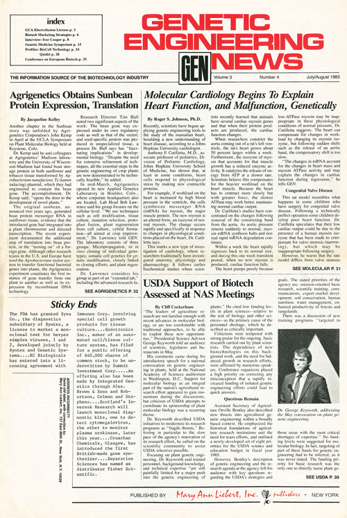
In our ongoing celebration of GEN’s 30th anniversary, we’ve reprinted an article from the July/August 1983 issue of GEN that discusses the coming of age of molecular cardiology. The article notes that scientists had begun to apply genetic engineering technology to obtain a clearer picture of heart problems. For example, the new paradigm looked at the role of myosin in human heart disease and at what types of molecular mechanisms underlying heart issues could be described. RNA studies and the development of rat models simulating human heart disorders were key components in the transition to the molecular way of looking at health issues.
The article points out that in 1983 there were only six laboratories around the world that had adopted the molecular approach. There are probably now hundreds if not thousands of labs that utilize the technique.
—John Sterling, Editor in Chief
“As Seen in GEN—Flashback” Volume 3, Number 4, July/August 1983
Molecular Cardiology Begins to Explain Heart Function, and Malfunction, Genetically
By Roger S. Johnson, Ph.D.
Recently, scientists have begun applying genetic engineering tools to the study of the mammalian heart, heralding a new understanding of heart disease, according to Johns Hopkins University cardiologist.
Anthony F. Cutilleta, M.D., associate professor of pediatrics, Division of Pediatric Cardiology, Johns Hopkins University School of Medicine, has shown that, at least in some conditions, heart cells respond to physiological stress by making new contractile proteins.
For example, if workload on the heart is increased by high blood pressure in the ventricle, the cells synthesize messenger RNA (mRNA) for myosin, the major muscle protein. The new myosin is an altered form, an isozyme of normal myosin. The change occurs rapidly and specifically in response to changes in physiological conditions placed on the heart, Dr. Cutilletta says.
This marks a new type of investigation of cardiology, where researchers traditionally have investigated anatomy, physiology and pharmacology. It follows earlier biochemical studies where scientists recently learned that animals have several cardiac myosin genes and that when their protein products are produced, the cardiac function changes.
When researchers constrict the aorta coming out of a rat’s left ventricle, the rat’s heart grows about 50 percent larger within a week. Furthermore, the isozyme of myosin that accounts for that muscle growth has a reduced enzyme activity. It catalyzes the release of energy from ATP at a slower rate. Scientists believe this compensates for the heavier workload on the heart muscle. Because the heart has to contract more slowly but with greater force, the slower ATPase may work better, maintaining normal cardiac output.
Dr. Cutilletta’s studies have concentrated on the changes following removal of the constricting band from the aorta. As aortic pressure returns suddenly to normal, myosin mRNA synthesis halts and normal rapid mRNA degradation continues.
Within a week the heart rapidly regresses back to its normal size, and during this one week transition period, when no new myosin is produced, cardiac function falls.
The heart pumps poorly because low-ATPase myosin may be inappropriate in these physiological conditions of normal pressure, Dr. Cutilletta suggests. The heart can compensate for changes in workload by changing its myosin isozyme, but following sudden shifts such as the release of an aortic band, the compensatory shift is too slow.
“The changes in mRNA account for the changes in heart mass and myosin ATPase activity and may explain the changes in cardiac function during regression,” he tells GEN.

Congenital Valve Disease
This rat model resembles what happens in some children who have surgery for congenital valve disease. Following a technically perfect operation some children develop poor heart function. Dr. Cutilletta speculates that their low cardiac output could be due to the presence of a human myosin isozyme that has been made to compensate for valve stenosis (narrowing), but which may be inappropriate following surgery.
However, he warns that the rat model differs from valve stenosis because it is initiated rapidly and because the aorta is banded above the coronary arteries, increasing their blood pressure and possibly their blood flow.
Similarly, Dr. Cutilletta cautiously evaluates the significance of his rat model study to human cardiac diseases. He suggests that certain cardiomyopathies could be caused by changes in either myosin or other proteins such as actin, troponin or tropomyosin, which regulate contractility.
“We’re opening a new area—molecular cardiology. At present this new discipline is led by about six labs throughout the world,” he says.
“The big impact is going to come when physiological mechanisms of the heart can be explained on a molecular level,” Dr. Cutilletta says. “The cardiac cell changes. In spite of being a highly differentiated cell, it doesn’t sit there contracting under the same conditions for the rest of its life. It adapts specifically and rapidly to inducible changes due to changes in pressure, for example.”
First, molecular cardiologists probably will start to apply these tools to understanding human cardiomyopathies, he predicts. Scientists do not know what causes most of these diseases, but they can apply these techniques and others to be developed to determine if hereditary and acquired cardiomyopathies result from changes in contractile proteins.
Once scientists develop a better understanding of how changes in particular contractile proteins regulate cardiac function, cardiologists may be able to use this information to diagnose and treat heart disease patients. For example some scientists have reported that following a heart attack, in which some cells die, the undamaged, neighboring cells hypertrophy. Dr. Cutilletta suspects they might be producing a different myosin.
Diagnosis for individual patients probably will be done by different, more rapid methods such as monoclonal antibodies, Dr. Cutilletta says.
First, however, researchers have to show that the type of myosin plays an important role in human heart disease and understand the mechanisms of this process.
This knowledge of molecular controls of the heart’s function will help cardiologists develop better therapeutic approaches, Dr. Cutilletta says. Applying the tools of genetic engineering to such a complex tissue as the heart is difficult but promises tremendous rewards.


