
July 1, 2015 (Vol. 35, No. 13)
Vicki Glaser Writer GEN
Next-Generation Sequencing Is Increasingly Making Headway in Clinical Medicine for Diagnostic and Prognostic Purposes
It’s early days, but next-generation sequencing (NGS) is increasingly making headway in clinical medicine for diagnostic and prognostic purposes, for guiding disease management, and for monitoring treatment response.
Progress is particularly striking in cancer, where an NGS technology called RNA sequencing (RNA-Seq) is being used to sequence the transcriptome and decode the mechanisms and pathways by which noncoding RNA regulates gene expression. By explicating the role of noncoding RNA in tumorigenesis, RNA-Seq is creating new opportunities for biomarker and drug target discovery.
Emerging data reveal the complexity of the various types of noncoding RNA elements—microRNAs (miRNAs), long noncoding RNAs (lncRNAs), circular RNAs (circRNAs)—and how they function and interact to maintain finely tuned post-transcriptional regulatory control over transcript abundance. As studies have shown, changes in the levels of miRNAs, for example, can lead to transcript dysregulation associated with cancer.
Novel molecular tools and techniques are being developed to improve the ability to profile the transcriptome and detect, quantify, and analyze noncoding RNA to study cancer biology and pursue new therapeutic targets for cancer. And multiplexed approaches that combine genomic and transcriptomic information are revealing the value added by integrating these datasets to search for cancer-related mutations in tumor samples.
The application of NGS to guide therapeutic decision-making in patients with advanced treatment-refractory cancers was discussed at a meeting recently held in Philadelphia by the American Association of Cancer Researchers (AACR).
A poster, “Implementation of CLIA Enabled Integrated Whole Genome (WGS)/Exome (WES)/Transcriptome (RNAseq) Next-Gen Sequencing to Identify Therapeutically Relevant Targets in Advanced Cancer Patients,” was presented by first author Mitesh Borad, M.D., a hematologist at the Mayo Clinic Cancer Center in Arizona, and colleagues.
The researchers described a workflow in which nucleic acids were extracted from tumor or bone marrow biopsies and used to perform WGS, WES, and RNA-Seq. Based on the sequencing data, the researchers prioritized therapeutic targets, which were then CLIA validated using Sanger sequencing. They identified one to two therapeutically targetable mutations per case in 60% of cases. Genomic targeted treatment was delivered to 13 patients, and preliminary clinical efficacy was reported in 5 patients.
In one example, a patient with an ERRFI1 mutation had a partial response to treatment with erlotinib. Citing the value of an integrated genomic/exomic/transcriptomic approach, the researchers reported that while the ERRFI1 allele was present in only 11% of the DNA reads, the mutant transcript was found in 82% of the RNA-Seq reads.
In another example, the researchers described a case in which whole exome sequencing and RNA-Seq data led to the identification of an FGFR2-MGEA5 fusion that was prioritized as a drug target. Tumor shrinkage was documented six weeks after treatment with the FGFR inhibitor panatinib.
Probing the Human Transcriptome
In another poster, “Characterization of the Human miRNA-ome Reveals Numerous Novel, Tissue-Specific miRNAs that are Implicated in Cancer Biology,” Eric Londin, Ph.D., Phillipe Loher, Ph.D., and Isidore Rigoutsos, Ph.D., of Sidney Kimmel College of Medicine at Thomas Jefferson University described a large collection of novel human miRNAs.
Processing of 1,323 deep sequencing (RNA-Seq) samples from 13 human tissues relied on the miRdeep2 algorithm to identify precursor and mature miRNAs. The sequencing process eliminated known miRNA structures, poorly expressed mature miRNAs (false discovery rate < 0.05), and miRNAs that intersected with snRNAs, tRNAs, or rRNAs.
The poster reported 3,707 newly identified mature miRNAs that are primate-specific. The sequence for the genomic loci of most of these were absent from mouse, Drosphila, or worm genomes. Therefore, the function of these miRNAs and their potential role in cancer could not be studied using mouse models of disease, for example.
The authors also reported tissue-specific expression for the novel miRNAs across a range of tissue types. And they presented data illustrating differential expression of the newly identified miRNAs among three cancer sample types—breast cancer, pancreatic cancer, and uveal melanoma.
Ling Lin, Ph.D., a clinical genomics R&D scientist at Dana-Farber Cancer Institute, was the first author of the poster “Targeted RNA Sequencing Improves Transcript Analysis in Cancer Samples.” In this presentation, Dr. Lin and colleagues stated that RNA-Seq can yield information on sequence, splicing (various transcripts), rearrangements, and expression levels (transcript abundance). According to the authors, the use of deep sequencing is necessitated by the complexity of the transcriptome and the wide dynamic range of expression levels, particularly for somatic mutation calling or fusion detection in low-abundance mRNAs.
For clinical applicability, the emphasis should be on the coding sequence, which is more likely to affect cell function. “To explore the feasibility of a more cost-effective method, we compared the performance of three different RNA-Seq methods: whole transcriptome, exome, and targeted RNA-Seq,” wrote the authors. To do so, they enriched the nucleic acid sample extracted from cancer cell lines and FFPE samples for 500 cancer-related genes, and depleted the RNA component of ribosomal RNA (rRNA).
Compared to whole transcriptome RNA-Seq, the authors observed that exome and targeted RNA-Seq demonstrated (1) higher coding exon coverage and multiplexing capability; (2) lower rRNA composition (reduced to 1%); (3) comparable gene abundance information; and (4) a high degree of alignment to coding exon regions in FFPE samples (over 90% compared to ~30% with the whole transcriptome method).
Overall, the researchers reported, targeted RNA-Seq increased coverage of the coding regions of genes of interest and so increased sensitivity of somatic mutation and fusion detection relative to whole transcriptome RNA-Seq. Relative gene-expression levels were preserved with targeted RNA-Seq.
Noncoding RNA is “the dark matter of the genome,” said Pier Paolo Pandolfi, M.D., a research geneticist with appointments at Beth Israel Deaconess Medical Center and Harvard Medical School. Dr. Pandolfi described work to identify, characterize, and functionalize the various types of RNA entities, as well as efforts to understand how these entities regulate gene expression and influence tumor formation and progression.
In his presentation, “In Vivo Analysis of Linear and Circular ceRNAs in Tumorigenesis,” Dr. Pandolfi said that “the math doesn’t add up.” While 70% of the genome is transcribed, only 2% of the RNA generated is mRNA that encodes protein; other linear RNA species are currently estimated at more than 3,000 miRNAs, 38,000 lncRNAs, and 20,000 pseudogenes. More recently, circular RNA elements have been identified, and the number of these new entities, noted Dr. Pandolfi, is “exploding,” with more than 100,000 already identified. Their relevance in cancer is garnering increasing attention.
Based on the evolving view of how miRNA networks operate and what Dr. Pandolfi calls the “new RNA code”—the language of competing endogenous RNAs (ceRNAs)—he envisions new opportunities for the discovery of novel cancer therapeutics when “we can predict which RNAs talk to which RNAs.” MicroRNAs play an important role in the life cycle of mRNA, and increased or decreased levels of specific miRNAs have been linked to cancer.
The theory behind the ceRNA language holds that any form of RNA, coding or noncoding, can act as a ceRNA, competing for miRNAs, which can be used for communication. If there is too little or too much miRNA, then communication between the various ceRNAs will be disrupted. According to Dr. Pandolfi, circRNAs, which are the most abundant RNA species in cells, “can modulate the crosstalk between linear ceRNAs by sequestering miRNAs in excess.”
In cancer, circRNAs have been shown to act as tumor suppressors in vitro. Recent in vivo studies suggest that the interplay between linear and circular RNAs contributes to tumorigenesis.
Ultimately, as researchers focused on understanding tumor biology and on new drug discovery in cancer explore how RNA elements interact within and between interconnected networks, they will look more closely at the composition of individual RNAs. However, “before we even begin to sequence noncoding RNAs, we need to determine systematically which RNAs are present and when,” advises Dr. Pandolfi.

Exiqon recently announced the launch of a microRNA sequencing service that includes RNA isolation, library preparation, sequencing, data analysis, and biological interpretation. The company says that it can provide high-quality results from samples with low RNA content, such as serum/plasma and other biofluids. In addition, it can deliver a final report just 4–6 weeks after receiving a sample.
Vendor Offerings
In the exhibit hall at the AACR meeting, vendors introduced new products, technologies, and services to improve and accelerate the sequencing, analysis, and expression profiling of all forms of coding and noncoding RNAs.
Arraystar presented its human and mouse circRNA microarrays, which it developed to study circular RNA expression and its role in human disease through the regulation of microRNAs. These microarrays use RNase R and circular junction specific probes to distinguish between circular and linear RNAs with high specificity.
Exiqon launched its microRNA sequencing service for serum and plasma samples at AACR. Using proprietary RNA isolation and microRNA library preparation protocols, Exiqon starts with a 500 µL serum or plasma sample and delivers sequence data in four to six weeks.
Thermo Fisher Scientifics’ new Oncomine Focus Assay is a library prep system that enables concurrent analysis of multiple DNA and RNA biomarkers in solid tumor samples using NGS on the company’s Ion PGM platform. The assay allows for the detection of more than 1,000 variants in 52 genes selected based on information in the Oncomine Knowledgebase. Each assay requires two or three slides, each containing 10 ng of nucleic acids derived from FFPE solid tumor samples.
Advanced Cell Diagnostics described its RNAscope RNA detection technology as a complementary tool to NGS, microarray-based expression analysis, and immunohistochemistry methods. The company’s biomarker detection and quantification approach is based on a multiplexed fluorescent and chromogenic in situ hybridization platform capable of single-molecule sensitivity.
The recently introduced NeoPrep Library Prep System from Illumina can process 16 RNA or DNA samples at a time in a 7.5-hour run for DNA and 10.5 hours for RNA that requires 30 minutes of hands-on time. Featuring digital microfluidic technology, the NeoPrep performs all sample processing on a self-contained library card. The TruSeq Stranded mRNA Library Prep Kit for NeoPrep requires 25–100 ng of input RNA to generate libraries for sequencing on an Illumina sequencing platform.
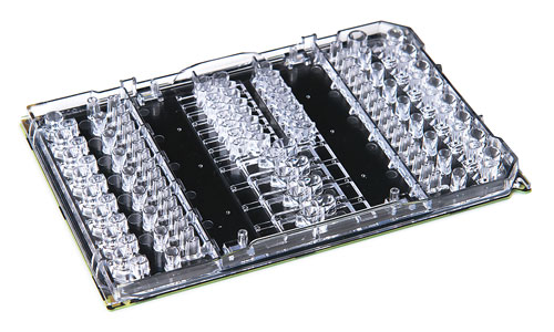
Illumina’s Neoprep Library Card, a tightly con-trolled environment for digital microfluidics, manipulates droplets to carry out library prep processes such as quantification and normalization.


