
February 1, 2011 (Vol. 31, No. 3)
Vicki Glaser Writer GEN
From Single Cell to Whole Organism, Change Is Under Way in Numerous Laboratories
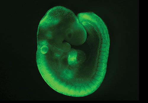
At the American Society for Cell Biology’s annual meeting last month, Kat Hadjantonakis, Ph.D., an associate member of the Sloan-Kettering Institute, spoke about imaging live-cell dynamics in mouse embryos. She described imaging experiments designed to shed light on cell lineage differentiation and morphogenesis during mouse development in utero.
Researchers can study embryo maturation in situ with the use of optical probes and techniques such as gene targeting to introduce into a fertilized egg a gene that codes for a fluorescent fusion protein reporter.
Dr. Hadjantonakis’ group used a fusion reporter composed of the H2B histone protein and green fluorescent protein (GFP) to image blastocyst-stage embryos (the stage at which stem cells form), which are approximately 140 microns in diameter and contain about 150 cells. The H2B-GFP mouse strains express the reporter in endodermal cells.
The group found that the inner cell mass of the blastocyst comprises two cell lineages in a random fashion (the pluripotent epiblast and the primitive endoderm), “suggesting plasticity in lineage specification,” Dr. Hadjantonakis reported. The embryos will ultimately eliminate via apoptosis those cells that do not undergo proper lineage specification, she added.
Platelet-derived growth factor receptor alpha (PDGFRα) can be used as a marker of the primitive endoderm lineage and its derivatives. Dr. Hadjantonakis introduced the H2B-GFP fusion reporter into blastocysts under the control of PDGFRα regulatory elements. Live imaging of the embryos led to the conclusion that PDGF signaling is essential for the establishment and expansion of the primitive endoderm lineage.
Dr. Hadjantonakis also showed how laser confocal microscopy can be used to achieve single-cell resolution of an embryo. Combined with tissue-specific fluorescent-labeling techniques, it is possible to visualize and distinguish between cells of the endoderm, ectoderm, and mesoderm as the embryo develops, and to demonstrate tissue-specific expression and study cell signaling at the level of a single cell. “We are moving to single-cell resolution data,” she explained.
In addition, she described how photoconvertible probes can be used to visualize cell population dynamics and follow a group of cells over time, to study, for example, mouse embryo morphogenesis and determine cell fate. Dr. Hadjantonakis reported that her group is exploring the combination of photoconversion and subcellular localization to enable single-cell precision imaging.
Photoconversion protein monomers typically have a half-life in vivo of 48 to 52 hours, depending on the tissue in which they are expressed. Any sensor protein used for intravital imaging must be thoroughly evaluated to determine its potential for cytotoxic effects such as cell aggregation.
In the future, multiplexing techniques will rely on fluorescent reporters for differential staining of the nucleus and cytoplasm. Intravital imaging can then be used to gain additional insights into the pathways and changes associated with cell morphology, cell division, and cell death. The ability to study cell activity and viability at the single cell level in vivo could have important implications in drug discovery for understanding the effects of experimental compounds.
A sensitive and bright single-cell resolution live imaging reporter of Wnt/ß-catenin signaling in the mouse. [Sloan-Kettering Institute]
Cell Dynamics
John Condeelis, Ph.D., professor and co-chair of the department of anatomy and structural biology at Albert Einstein College of Medicine of Yeshiva University, spoke about the application of intravital multiphoton microscopy and photoconversion technology to define cell dynamics and cell-cell interactions that drive normal morphogenesis and metastatic tumor progression in the mouse mammary gland. A clearer picture of the early stages of metastatic behavior could lead to the identification of novel targets for chemotherapeutic drug development.
As single-cell movement is the dominant mode of metastasis, technology is needed to enable the study of cell motility and the tumor microenvironment at single-cell resolution. The microenvironment plays a major role in determining the gene-expression profile and migratory phenotype of tumor cells, he explained.
The transparent nature of embryos facilitates in vivo imaging studies. To image adult organisms—solid tissues and tumors in particular—and to study the behavior of single cells, Dr. Condeelis utilizes multiphoton imaging technology, which allows for visualization “to depths 10x to 30x greater than one photon excitation.”
New microscope designs enable scanning of multiple photons simultaneously using probes that fluoresce at different wavelengths across the spectrum detectable by a particular imaging device. Photoconvertible proteins are used to follow cell movement and morphological changes over time. Novel exotic fluorophores and new generations of photoconvertible proteins are contributing to rapid advances in the field.
To facilitate repeated imaging of mammary tumors in individual mice, Dr. Condeelis’ group developed a technique in which they suture a “window” in place of the epidermal layer of skin that overlies the mammary gland. He showed in vivo images obtained after labeling a subpopulation of mammary cells using photoconvertible probes.
The images capture the early stages of metastasis, in which streams of cells that have made the decision to migrate utilize extracellular matrix fibers to access the blood supply. The images point to a key difference between the microenvironment in which cell intravasation occurs versus where it does not: the presence of perivascular clusters of invasive, tumor-associated macrophages, which serve as a source of chemotactic growth factors that mediate tumor cell-macrophage interaction. He described this population of macrophages as “doormen” that create an entry point and support the efficient invasion of tumor cells into the bloodstream.
Similar types of choreographed co-migratory events have been described during normal embryonic development and mammary gland morphogenesis, and these rely on many of the same signaling, steering, and invasive pathways that drive metastatic tumor cell behavior.
Drug Effects
Ralph Weissleder, M.D., Ph.D., professor, department of radiology at Harvard Medical School and director, Center for Molecular Imaging Research at Massachusetts General Hospital, described his research on in vivo imaging of drug effects and the results of experiments designed to understand the biological basis of tumor cell interaction with host cells.
A primary goal of this work is to translate the use of intravital microscopy and injectable fluorescent probes from the research laboratory to the clinic for the purpose of assessing the effects of chemotherapeutic agents on tumor and normal cells and identifying more targeted cancer therapies.
Advances in the adaptation of cell-based assays to intravital imaging in the mouse have demonstrated fundamental differences between what is seen in vitro versus in vivo. Dr. Weissleder emphasized three specific benefits that intravital microscopy offers cancer researchers: the potential to observe physiological responses, to study heterogeneity within populations of cancer cells or between cellular processes, and to image drug responses. How best to achieve these benefits—what cell and tumor types to study, how long to image and follow cell populations, and what imaging parameters are optimal, for example—are all questions that require further study.
Tumors can be imaged in situ by applying various techniques including the use of “stick” lenses, or microscope objectives that enable “keyhole” imaging, as well as imaging modalities such as laser-scanning confocal or multiphoton microscopy. Studying drug responses in tumors requires high spatial resolution to observe nuclear phenotypes and detect nuclear division, explained Dr. Weissleder.
Whole tumor imaging can be achieved at high resolution by imaging tumor slices and then combining the individual images—a process called “image stitching.”
As drug responses evolve over time and may be heterogeneous across tumor cell populations, the ability to do time-lapse imaging and sequential studies in the same animal over multiple time points is important. This presents a particular challenge as tumors are dynamic structures; it requires the development of methods and technologies that will allow researchers to image the same group of cells in vivo over a period of hours to days.
Cost can be a significant barrier to entry into the emerging field of intravital imaging. While microscopy systems are available in a range of complexity, capabilities, and costs, with personal confocal microscopes available for as low as $100,000, and multiphoton imaging systems for $200,000, instrument costs are only one component of the overall expense. Additional infrastructure needs to build and maintain small animal and surgical facilities must also be considered.


